5LM1

Crystal Structure of HD-PTP phosphatase in complex with UBAP1


Experimental Data Snapshot

  • Method: X-RAY DIFFRACTION
  • Resolution: 2.55 Å
  • R-Value Free: 0.254 
  • R-Value Work: 0.213 
  • R-Value Observed: 0.215 

Starting Model: other
View more details

wwPDB Validation   3D Report Full Report


This is version 1.2 of the entry. See complete history


Literature

Structural Basis for Selective Interaction between the ESCRT Regulator HD-PTP and UBAP1.

Gahloth, D.Levy, C.Heaven, G.Stefani, F.Wunderley, L.Mould, P.Cliff, M.J.Bella, J.Fielding, A.J.Woodman, P.Tabernero, L.

(2016) Structure 24: 2115-2126

  • DOI: https://doi.org/10.1016/j.str.2016.10.006
  • Primary Citation of Related Structures:  
    5LM1, 5LM2

  • PubMed Abstract: 

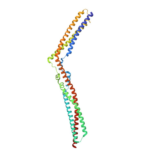
    Endosomal sorting complexes required for transport (ESCRTs) are essential for ubiquitin-dependent degradation of mitogenic receptors, a process often compromised in cancer pathologies. Sorting of ubiquinated receptors via ESCRTs is controlled by the tumor suppressor phosphatase HD-PTP. The specific interaction between HD-PTP and the ESCRT-I subunit UBAP1 is critical for degradation of growth factor receptors and integrins. Here, we present the structural characterization by X-ray crystallography and double electron-electron resonance spectroscopy of the coiled-coil domain of HD-PTP and its complex with UBAP1. The coiled-coil domain adopts an unexpected open and rigid conformation that contrasts with the closed and flexible coiled-coil domain of the related ESCRT regulator Alix. The HD-PTP:UBAP1 structure identifies the molecular determinants of the interaction and provides a molecular basis for the specific functional cooperation between HD-PTP and UBAP1. Our findings provide insights into the molecular mechanisms of regulation of ESCRT pathways that could be relevant to anticancer therapies.


  • Organizational Affiliation

    School of Biological Sciences, Faculty of Biology, Medicine and Health, University of Manchester, Manchester M13 9PT, UK.


Macromolecules
Find similar proteins by:  (by identity cutoff)  |  3D Structure
Entity ID: 1
MoleculeChains Sequence LengthOrganismDetailsImage
Tyrosine-protein phosphatase non-receptor type 23352Homo sapiensMutation(s): 0 
Gene Names: PTPN23KIAA1471
EC: 3.1.3.48
UniProt & NIH Common Fund Data Resources
Find proteins for Q9H3S7 (Homo sapiens)
Explore Q9H3S7 
Go to UniProtKB:  Q9H3S7
PHAROS:  Q9H3S7
GTEx:  ENSG00000076201 
Entity Groups  
Sequence Clusters30% Identity50% Identity70% Identity90% Identity95% Identity100% Identity
UniProt GroupQ9H3S7
Sequence Annotations
Expand
  • Reference Sequence

Find similar proteins by:  Sequence   |   3D Structure  

Entity ID: 2
MoleculeChains Sequence LengthOrganismDetailsImage
UBAP-120Homo sapiensMutation(s): 0 
UniProt & NIH Common Fund Data Resources
Find proteins for Q9NZ09 (Homo sapiens)
Explore Q9NZ09 
Go to UniProtKB:  Q9NZ09
PHAROS:  Q9NZ09
GTEx:  ENSG00000165006 
Entity Groups  
Sequence Clusters30% Identity50% Identity70% Identity90% Identity95% Identity100% Identity
UniProt GroupQ9NZ09
Sequence Annotations
Expand
  • Reference Sequence
Experimental Data & Validation

Experimental Data

  • Method: X-RAY DIFFRACTION
  • Resolution: 2.55 Å
  • R-Value Free: 0.254 
  • R-Value Work: 0.213 
  • R-Value Observed: 0.215 
  • Space Group: P 21 21 21
Unit Cell:
Length ( Å )Angle ( ˚ )
a = 48.93α = 90
b = 93.27β = 90
c = 102.22γ = 90
Software Package:
Software NamePurpose
PHENIXrefinement
xia2data reduction
xia2data scaling
PHASERphasing

Structure Validation

View Full Validation Report


Entry History 

Deposition Data

  • Released Date: 2016-11-30 
  • Deposition Author(s): Levy, C.

Revision History  (Full details and data files)

  • Version 1.0: 2016-11-30
    Type: Initial release
  • Version 1.1: 2016-12-14
    Changes: Database references
  • Version 1.2: 2024-05-01
    Changes: Data collection, Database references, Refinement description