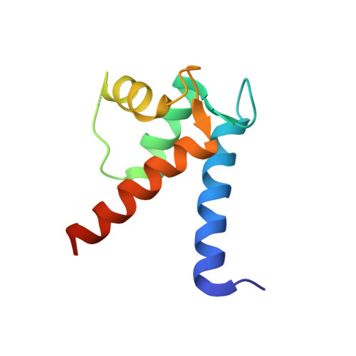
X-ray crystal structure of human calcium-bound S100A1.
Melville, Z., Aligholizadeh, E., McKnight, L.E., Weber, D.J., Pozharski, E., Weber, D.J.(2017) Acta Crystallogr F Struct Biol Commun 73: 215-221
- PubMed: 28368280
- DOI: https://doi.org/10.1107/S2053230X17003983
- Primary Citation of Related Structures:
5K89 - PubMed Abstract:
S100A1 is a member of the S100 family of Ca 2+ -binding proteins and regulates several cellular processes, including those involved in Ca 2+ signaling and cardiac and skeletal muscle function. In Alzheimer's disease, brain S100A1 is overexpressed and gives rise to disease pathologies, making it a potential therapeutic target. The 2.25 Å resolution crystal structure of Ca 2+ -S100A1 is solved here and is compared with the structures of other S100 proteins, most notably S100B, which is a highly homologous S100-family member that is implicated in the progression of malignant melanoma. The observed structural differences in S100A1 versus S100B provide insights regarding target protein-binding specificity and for targeting these two S100 proteins in human diseases using structure-based drug-design approaches.
Organizational Affiliation:
Center for Biomolecular Therapeutics, Department of Biochemistry and Molecular Biology, University of Maryland Baltimore, 108 North Greene Street, Baltimore, MD 21201, USA.