5JSF

Crystal structure of 17beta-hydroxysteroid dehydrogenase 14 S205 variant in complex with NAD.


Experimental Data Snapshot

  • Method: X-RAY DIFFRACTION
  • Resolution: 1.84 Å
  • R-Value Free: 0.187 
  • R-Value Work: 0.165 
  • R-Value Observed: 0.166 

Starting Model: experimental
View more details

wwPDB Validation   3D Report Full Report


Ligand Structure Quality Assessment 


This is version 1.3 of the entry. See complete history


Literature

New Insights into Human 17 beta-Hydroxysteroid Dehydrogenase Type 14: First Crystal Structures in Complex with a Steroidal Ligand and with a Potent Nonsteroidal Inhibitor.

Bertoletti, N.Braun, F.Lepage, M.Moller, G.Adamski, J.Heine, A.Klebe, G.Marchais-Oberwinkler, S.

(2016) J Med Chem 59: 6961-6967

  • DOI: https://doi.org/10.1021/acs.jmedchem.6b00293
  • Primary Citation of Related Structures:  
    5HS6, 5ICM, 5ICS, 5JS6, 5JSF

  • PubMed Abstract: 

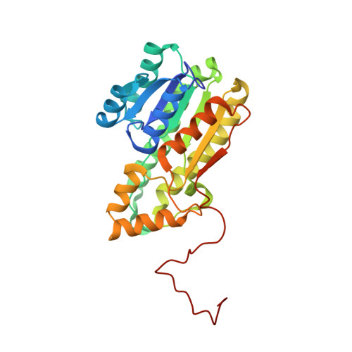
    17β-HSD14 is a SDR enzyme able to oxidize estradiol and 5-androstenediol using NAD(+). We determined the crystal structure of this human enzyme as the holo form and as ternary complexes with estrone and with the first potent, nonsteroidal inhibitor. The structures reveal a conical, rather large and lipophilic binding site and are the starting point for structure-based inhibitor design. The two natural variants (S205 and T205) were characterized and adopt a similar structure.


  • Organizational Affiliation

    Institute for Pharmaceutical Chemistry, Philipps University Marburg , 35037 Marburg, Germany.


Macromolecules
Find similar proteins by:  (by identity cutoff)  |  3D Structure
Entity ID: 1
MoleculeChains Sequence LengthOrganismDetailsImage
17-beta-hydroxysteroid dehydrogenase 14274Homo sapiensMutation(s): 0 
Gene Names: HSD17B14DHRS10SDR3SDR47C1UNQ502/PRO474
EC: 1.1.1
UniProt & NIH Common Fund Data Resources
Find proteins for Q9BPX1 (Homo sapiens)
Explore Q9BPX1 
Go to UniProtKB:  Q9BPX1
PHAROS:  Q9BPX1
GTEx:  ENSG00000087076 
Entity Groups  
Sequence Clusters30% Identity50% Identity70% Identity90% Identity95% Identity100% Identity
UniProt GroupQ9BPX1
Sequence Annotations
Expand
  • Reference Sequence
Experimental Data & Validation

Experimental Data

  • Method: X-RAY DIFFRACTION
  • Resolution: 1.84 Å
  • R-Value Free: 0.187 
  • R-Value Work: 0.165 
  • R-Value Observed: 0.166 
  • Space Group: I 2 3
Unit Cell:
Length ( Å )Angle ( ˚ )
a = 130.256α = 90
b = 130.256β = 90
c = 130.256γ = 90
Software Package:
Software NamePurpose
PHENIXrefinement
XDSdata reduction
XDSdata scaling
PHASERphasing

Structure Validation

View Full Validation Report


Ligand Structure Quality Assessment 


Entry History & Funding Information

Deposition Data


Funding OrganizationLocationGrant Number
German Research FoundationGermanyMA-5287/1-1
German Research FoundationGermanyKL-1204/15-1

Revision History  (Full details and data files)

  • Version 1.0: 2016-07-13
    Type: Initial release
  • Version 1.1: 2016-08-10
    Changes: Database references
  • Version 1.2: 2017-09-06
    Changes: Author supporting evidence
  • Version 1.3: 2024-01-10
    Changes: Data collection, Database references, Refinement description