5JG6

APC11-Ubv shows role of noncovalent RING-Ubiquitin interactions in processive multiubiquitination and Ubiquitin chain elongation by APC/C

  • Classification: CELL CYCLE
  • Organism(s): Homo sapiens
  • Expression System: Escherichia coli
  • Mutation(s): No 

  • Deposited: 2016-04-19 Released: 2016-06-15 
  • Deposition Author(s): Brown, N.G., Zhang, W., Yu, S., Miller, D.J., Sidhu, S.S., Schulman, B.A.
  • Funding Organization(s): National Institutes of Health/National Institute of General Medical Sciences (NIH/NIGMS), National Institutes of Health/National Cancer Institute (NIH/NCI), St. Jude Children's Research Hospital (ALSAC), Howard Hughes Medical Institute (HHMI), Canadian Institutes of Health Research (CIHR), Leukemia & Lymphoma Society, Jane Coffin Childs Memorial Fund

Experimental Data Snapshot

  • Method: X-RAY DIFFRACTION
  • Resolution: 2.00 Å
  • R-Value Free: 0.220 
  • R-Value Work: 0.185 
  • R-Value Observed: 0.187 

Starting Models: experimental
View more details

wwPDB Validation   3D Report Full Report


This is version 1.5 of the entry. See complete history


Literature

Dual RING E3 Architectures Regulate Multiubiquitination and Ubiquitin Chain Elongation by APC/C.

Brown, N.G.VanderLinden, R.Watson, E.R.Weissmann, F.Ordureau, A.Wu, K.P.Zhang, W.Yu, S.Mercredi, P.Y.Harrison, J.S.Davidson, I.F.Qiao, R.Lu, Y.Dube, P.Brunner, M.R.Grace, C.R.Miller, D.J.Haselbach, D.Jarvis, M.A.Yamaguchi, M.Yanishevski, D.Petzold, G.Sidhu, S.S.Kuhlman, B.Kirschner, M.W.Harper, J.W.Peters, J.M.Stark, H.Schulman, B.A.

(2016) Cell 165: 1440-1453

  • DOI: https://doi.org/10.1016/j.cell.2016.05.037
  • Primary Citation of Related Structures:  
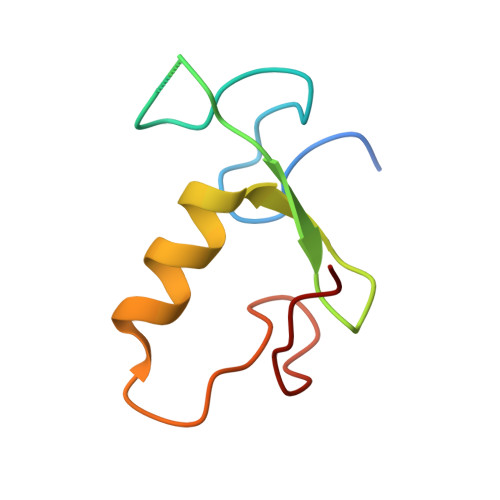
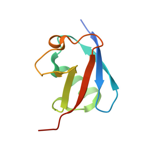
    5JG6, 5L9T, 5L9U

  • PubMed Abstract: 

    Protein ubiquitination involves E1, E2, and E3 trienzyme cascades. E2 and RING E3 enzymes often collaborate to first prime a substrate with a single ubiquitin (UB) and then achieve different forms of polyubiquitination: multiubiquitination of several sites and elongation of linkage-specific UB chains. Here, cryo-EM and biochemistry show that the human E3 anaphase-promoting complex/cyclosome (APC/C) and its two partner E2s, UBE2C (aka UBCH10) and UBE2S, adopt specialized catalytic architectures for these two distinct forms of polyubiquitination. The APC/C RING constrains UBE2C proximal to a substrate and simultaneously binds a substrate-linked UB to drive processive multiubiquitination. Alternatively, during UB chain elongation, the RING does not bind UBE2S but rather lures an evolving substrate-linked UB to UBE2S positioned through a cullin interaction to generate a Lys11-linked chain. Our findings define mechanisms of APC/C regulation, and establish principles by which specialized E3-E2-substrate-UB architectures control different forms of polyubiquitination.


  • Organizational Affiliation

    Department of Structural Biology, St. Jude Children's Research Hospital, Memphis, TN 38105, USA.


Macromolecules
Find similar proteins by:  (by identity cutoff)  |  3D Structure
Entity ID: 1
MoleculeChains Sequence LengthOrganismDetailsImage
Anaphase-promoting complex subunit 11
A, D
70Homo sapiensMutation(s): 0 
Gene Names: ANAPC11HSPC214
UniProt & NIH Common Fund Data Resources
Find proteins for Q9NYG5 (Homo sapiens)
Explore Q9NYG5 
Go to UniProtKB:  Q9NYG5
PHAROS:  Q9NYG5
GTEx:  ENSG00000141552 
Entity Groups  
Sequence Clusters30% Identity50% Identity70% Identity90% Identity95% Identity100% Identity
UniProt GroupQ9NYG5
Sequence Annotations
Expand
  • Reference Sequence
Find similar proteins by:  (by identity cutoff)  |  3D Structure
Entity ID: 2
MoleculeChains Sequence LengthOrganismDetailsImage
Polyubiquitin-B
B, C
84Homo sapiensMutation(s): 0 
Gene Names: UBB
UniProt & NIH Common Fund Data Resources
Find proteins for P0CG47 (Homo sapiens)
Explore P0CG47 
Go to UniProtKB:  P0CG47
GTEx:  ENSG00000170315 
Entity Groups  
Sequence Clusters30% Identity50% Identity70% Identity90% Identity95% Identity100% Identity
UniProt GroupP0CG47
Sequence Annotations
Expand
  • Reference Sequence
Experimental Data & Validation

Experimental Data

  • Method: X-RAY DIFFRACTION
  • Resolution: 2.00 Å
  • R-Value Free: 0.220 
  • R-Value Work: 0.185 
  • R-Value Observed: 0.187 
  • Space Group: C 1 2 1
Unit Cell:
Length ( Å )Angle ( ˚ )
a = 131.066α = 90
b = 35.193β = 120.42
c = 72.768γ = 90
Software Package:
Software NamePurpose
PHENIXrefinement
HKL-2000data reduction
HKL-2000data scaling
PHASERphasing

Structure Validation

View Full Validation Report


Entry History & Funding Information

Deposition Data


Funding OrganizationLocationGrant Number
National Institutes of Health/National Institute of General Medical Sciences (NIH/NIGMS)United StatesR37GM065930
National Institutes of Health/National Cancer Institute (NIH/NCI)United StatesP30CA021765
St. Jude Children's Research Hospital (ALSAC)United States--
Howard Hughes Medical Institute (HHMI)United States--
Canadian Institutes of Health Research (CIHR)CanadaMOP#111149
Canadian Institutes of Health Research (CIHR)Canada136956
Leukemia & Lymphoma SocietyUnited States--
Jane Coffin Childs Memorial FundUnited States--

Revision History  (Full details and data files)

  • Version 1.0: 2016-06-15
    Type: Initial release
  • Version 1.1: 2017-09-06
    Changes: Author supporting evidence, Derived calculations
  • Version 1.2: 2017-09-27
    Changes: Author supporting evidence
  • Version 1.3: 2019-11-20
    Changes: Author supporting evidence
  • Version 1.4: 2020-01-08
    Changes: Author supporting evidence
  • Version 1.5: 2023-09-27
    Changes: Data collection, Database references, Refinement description