5GAL

CRYSTAL STRUCTURE OF HUMAN GALECTIN-7 IN COMPLEX WITH N-ACETYLLACTOSAMINE


Experimental Data Snapshot

  • Method: X-RAY DIFFRACTION
  • Resolution: 2.00 Å
  • R-Value Free: 0.296 
  • R-Value Work: 0.213 
  • R-Value Observed: 0.213 

Starting Model: experimental
View more details

wwPDB Validation   3D Report Full Report


This is version 2.2 of the entry. See complete history


Literature

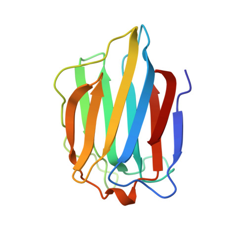
Structural basis for the recognition of carbohydrates by human galectin-7.

Leonidas, D.D.Vatzaki, E.H.Vorum, H.Celis, J.E.Madsen, P.Acharya, K.R.

(1998) Biochemistry 37: 13930-13940

  • DOI: https://doi.org/10.1021/bi981056x
  • Primary Citation of Related Structures:  
    1BKZ, 2GAL, 3GAL, 4GAL, 5GAL

  • PubMed Abstract: 

    Knowledge about carbohydrate recognition domains of galectins, formerly known as S-type animal lectins, is important in understanding their role(s) in cell-cell interactions. Here we report the crystal structure of human galectin-7 (hGal-7), in free form and in the presence of galactose, galactosamine, lactose, and N-acetyl-lactosamine at high resolution. This is the first structure of a galectin determined in both free and carbohydrate-bound forms. The structure shows a fold similar to that of the prototype galectins -1 and -2, but has greater similarity to a related galectin molecule, Gal-10. Even though the carbohydrate-binding residues are conserved, there are significant changes in this pocket due to shortening of a loop structure. The monomeric hGal-7 molecule exists as a dimer in the crystals, but adopts a packing arrangement considerably different from that of Gal-1 and Gal-2, which has implications for carbohydrate recognition.


  • Organizational Affiliation

    Department of Biology and Biochemistry, University of Bath, UK.


Macromolecules
Find similar proteins by:  (by identity cutoff)  |  3D Structure
Entity ID: 1
MoleculeChains Sequence LengthOrganismDetailsImage
GALECTIN-7
A, B
135Homo sapiensMutation(s): 0 
UniProt & NIH Common Fund Data Resources
Find proteins for P47929 (Homo sapiens)
Explore P47929 
Go to UniProtKB:  P47929
PHAROS:  P47929
Entity Groups  
Sequence Clusters30% Identity50% Identity70% Identity90% Identity95% Identity100% Identity
UniProt GroupP47929
Sequence Annotations
Expand
  • Reference Sequence
Oligosaccharides

Help

Entity ID: 2
MoleculeChains Length2D Diagram Glycosylation3D Interactions
beta-D-galactopyranose-(1-4)-2-acetamido-2-deoxy-beta-D-glucopyranose
C
2N/A
Glycosylation Resources
GlyTouCan:  G00055MO
GlyCosmos:  G00055MO
GlyGen:  G00055MO
Experimental Data & Validation

Experimental Data

  • Method: X-RAY DIFFRACTION
  • Resolution: 2.00 Å
  • R-Value Free: 0.296 
  • R-Value Work: 0.213 
  • R-Value Observed: 0.213 
  • Space Group: P 21 21 21
Unit Cell:
Length ( Å )Angle ( ˚ )
a = 54.23α = 90
b = 65.18β = 90
c = 73.39γ = 90
Software Package:
Software NamePurpose
X-PLORmodel building
X-PLORrefinement
DENZOdata reduction
SCALEPACKdata scaling
X-PLORphasing

Structure Validation

View Full Validation Report


Entry History 

Deposition Data

Revision History  (Full details and data files)

  • Version 1.0: 1998-11-04
    Type: Initial release
  • Version 1.1: 2008-03-25
    Changes: Version format compliance
  • Version 1.2: 2011-07-13
    Changes: Non-polymer description, Version format compliance
  • Version 2.0: 2020-07-29
    Type: Remediation
    Reason: Carbohydrate remediation
    Changes: Advisory, Atomic model, Data collection, Derived calculations, Other, Structure summary
  • Version 2.1: 2024-03-13
    Changes: Data collection, Database references, Structure summary
  • Version 2.2: 2024-04-03
    Changes: Refinement description