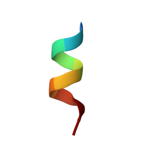
5G46

Ligand complex of RORg LBD


Experimental Data Snapshot

  • Method: X-RAY DIFFRACTION
  • Resolution: 1.76 Å
  • R-Value Free: 0.217 
  • R-Value Work: 0.186 
  • R-Value Observed: 0.187 

wwPDB Validation   3D Report Full Report


Ligand Structure Quality Assessment 


This is version 1.2 of the entry. See complete history


Literature

Fragment Screening of Rorgammat Using Cocktail Crystallography: Identification of Simultaneous Binding of Multiple Fragments.

Xue, Y.Guo, H.Hillertz, P.

(2016) ChemMedChem 11: 1881

  • DOI: https://doi.org/10.1002/cmdc.201600242
  • Primary Citation of Related Structures:  
    5G42, 5G43, 5G44, 5G45, 5G46

  • PubMed Abstract: 

    The retinoic-acid-related orphan receptor γ t (RORγt), as a master regulator of Th17 cell pathology, has become an attractive target for small-molecule drug discovery for the treatment of Th17-cell-related autoimmune diseases. A crystallographic fragment screening was carried out for RORγt using the ligand binding domain. An overall hit rate of 5.5 % was obtained by screening 384 compounds in 96 cocktails. Five distinct hotspots were identified, and four regions of anchoring polar interactions were observed. In addition, significant induced fit was found for the binding of several fragments. Strikingly, a simultaneous binding of three fragments was revealed which presents interesting features including π-π stacking, multiple hydrogen bonds to the protein, and significant induced fit. Overall, the results offer a complete mapping of the ligand binding pocket and provide valuable inspiration in structure-based design for RORγt lead generation and optimization. The crystallographic screening also resulted in fragment hits that bind at the surface away from the ligand binding pocket. This surface site is near the plausible dimer interface by analogy with other nuclear receptor systems, which can provide initial hints to explore alternative ways to modulate RORγt through protein-protein interactions.


  • Organizational Affiliation

    Discovery Sciences, Innovative Medicines and Early Development Biotech Unit, AstraZeneca Gothenburg, Pepparedsleden 1, 431 83, Mölndal, Sweden. yafeng.xue@astrazeneca.com.


Macromolecules
Find similar proteins by:  (by identity cutoff)  |  3D Structure
Entity ID: 1
MoleculeChains Sequence LengthOrganismDetailsImage
NUCLEAR RECEPTOR ROR-GAMMA266Homo sapiensMutation(s): 0 
UniProt & NIH Common Fund Data Resources
Find proteins for P51449 (Homo sapiens)
Explore P51449 
Go to UniProtKB:  P51449
PHAROS:  P51449
GTEx:  ENSG00000143365 
Entity Groups  
Sequence Clusters30% Identity50% Identity70% Identity90% Identity95% Identity100% Identity
UniProt GroupP51449
Sequence Annotations
Expand
  • Reference Sequence

Find similar proteins by:  Sequence   |   3D Structure  

Entity ID: 2
MoleculeChains Sequence LengthOrganismDetailsImage
RORGB [auth C]10Homo sapiensMutation(s): 0 
UniProt & NIH Common Fund Data Resources
Find proteins for Q15596 (Homo sapiens)
Explore Q15596 
Go to UniProtKB:  Q15596
PHAROS:  Q15596
GTEx:  ENSG00000140396 
Entity Groups  
Sequence Clusters30% Identity50% Identity70% Identity90% Identity95% Identity100% Identity
UniProt GroupQ15596
Sequence Annotations
Expand
  • Reference Sequence
Experimental Data & Validation

Experimental Data

  • Method: X-RAY DIFFRACTION
  • Resolution: 1.76 Å
  • R-Value Free: 0.217 
  • R-Value Work: 0.186 
  • R-Value Observed: 0.187 
  • Space Group: P 41 21 2
Unit Cell:
Length ( Å )Angle ( ˚ )
a = 61.96α = 90
b = 61.96β = 90
c = 154.57γ = 90
Software Package:
Software NamePurpose
BUSTERrefinement
XDSdata reduction
SCALAdata scaling

Structure Validation

View Full Validation Report


Ligand Structure Quality Assessment 


Entry History 

Deposition Data

Revision History  (Full details and data files)

  • Version 1.0: 2016-08-03
    Type: Initial release
  • Version 1.1: 2016-09-28
    Changes: Database references
  • Version 1.2: 2024-05-08
    Changes: Data collection, Database references, Derived calculations, Other