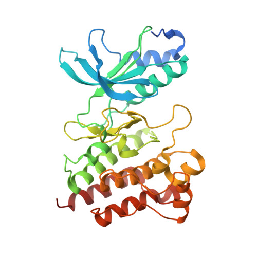
5DG5

CRYSTAL STRUCTURE OF THE TYROSINE KINASE DOMAIN OF THE HEPATOCYTE GROWTH FACTOR RECEPTOR C-MET IN COMPLEX WITH ALTIRATINIB ANALOG DP-4157


Experimental Data Snapshot

  • Method: X-RAY DIFFRACTION
  • Resolution: 2.60 Å
  • R-Value Free: 0.245 
  • R-Value Work: 0.204 
  • R-Value Observed: 0.206 

Starting Model: experimental
View more details

wwPDB Validation   3D Report Full Report


Ligand Structure Quality Assessment 


This is version 1.1 of the entry. See complete history


Literature

Altiratinib Inhibits Tumor Growth, Invasion, Angiogenesis, and Microenvironment-Mediated Drug Resistance via Balanced Inhibition of MET, TIE2, and VEGFR2.

Smith, B.D.Kaufman, M.D.Leary, C.B.Turner, B.A.Wise, S.C.Ahn, Y.M.Booth, R.J.Caldwell, T.M.Ensinger, C.L.Hood, M.M.Lu, W.P.Patt, T.W.Patt, W.C.Rutkoski, T.J.Samarakoon, T.Telikepalli, H.Vogeti, L.Vogeti, S.Yates, K.M.Chun, L.Stewart, L.J.Clare, M.Flynn, D.L.

(2015) Mol Cancer Ther 14: 2023-2034

  • DOI: https://doi.org/10.1158/1535-7163.MCT-14-1105
  • Primary Citation of Related Structures:  
    5DG5

  • PubMed Abstract: 

    Altiratinib (DCC-2701) was designed based on the rationale of engineering a single therapeutic agent able to address multiple hallmarks of cancer (1). Specifically, altiratinib inhibits not only mechanisms of tumor initiation and progression, but also drug resistance mechanisms in the tumor and microenvironment through balanced inhibition of MET, TIE2 (TEK), and VEGFR2 (KDR) kinases. This profile was achieved by optimizing binding into the switch control pocket of all three kinases, inducing type II inactive conformations. Altiratinib durably inhibits MET, both wild-type and mutated forms, in vitro and in vivo. Through its balanced inhibitory potency versus MET, TIE2, and VEGFR2, altiratinib provides an agent that inhibits three major evasive (re)vascularization and resistance pathways (HGF, ANG, and VEGF) and blocks tumor invasion and metastasis. Altiratinib exhibits properties amenable to oral administration and exhibits substantial blood-brain barrier penetration, an attribute of significance for eventual treatment of brain cancers and brain metastases.


  • Organizational Affiliation

    Deciphera Pharmaceuticals, Lawrence, Kansas.


Macromolecules
Find similar proteins by:  (by identity cutoff)  |  3D Structure
Entity ID: 1
MoleculeChains Sequence LengthOrganismDetailsImage
Hepatocyte growth factor receptor
A, B
318Homo sapiensMutation(s): 0 
Gene Names: MET
EC: 2.7.10.1
UniProt & NIH Common Fund Data Resources
Find proteins for P08581 (Homo sapiens)
Explore P08581 
Go to UniProtKB:  P08581
PHAROS:  P08581
GTEx:  ENSG00000105976 
Entity Groups  
Sequence Clusters30% Identity50% Identity70% Identity90% Identity95% Identity100% Identity
UniProt GroupP08581
Sequence Annotations
Expand
  • Reference Sequence
Small Molecules
Ligands 1 Unique
IDChains Name / Formula / InChI Key2D Diagram3D Interactions
5B4
Query on 5B4

Download Ideal Coordinates CCD File 
C [auth A],
D [auth B]
N-(2,5-difluoro-4-{[2-(1-methyl-1H-pyrazol-4-yl)pyridin-4-yl]oxy}phenyl)-N'-(4-fluorophenyl)cyclopropane-1,1-dicarboxam ide
C26 H20 F3 N5 O3
YLLMKENFMVEIDG-UHFFFAOYSA-N
Experimental Data & Validation

Experimental Data

  • Method: X-RAY DIFFRACTION
  • Resolution: 2.60 Å
  • R-Value Free: 0.245 
  • R-Value Work: 0.204 
  • R-Value Observed: 0.206 
  • Space Group: P 1 21 1
Unit Cell:
Length ( Å )Angle ( ˚ )
a = 37.13α = 90
b = 115.88β = 92.92
c = 90.16γ = 90
Software Package:
Software NamePurpose
XDSdata reduction
XSCALEdata scaling
PHASERphasing
Cootmodel building
REFMACrefinement

Structure Validation

View Full Validation Report


Ligand Structure Quality Assessment 


Entry History 

Revision History  (Full details and data files)

  • Version 1.0: 2016-08-31
    Type: Initial release
  • Version 1.1: 2023-09-27
    Changes: Data collection, Database references, Derived calculations, Refinement description, Structure summary