5CCL

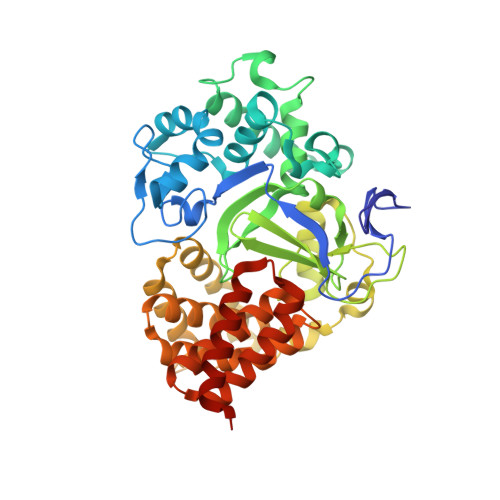
Crystal structure of SMYD3 with SAM and oxindole compound


Experimental Data Snapshot

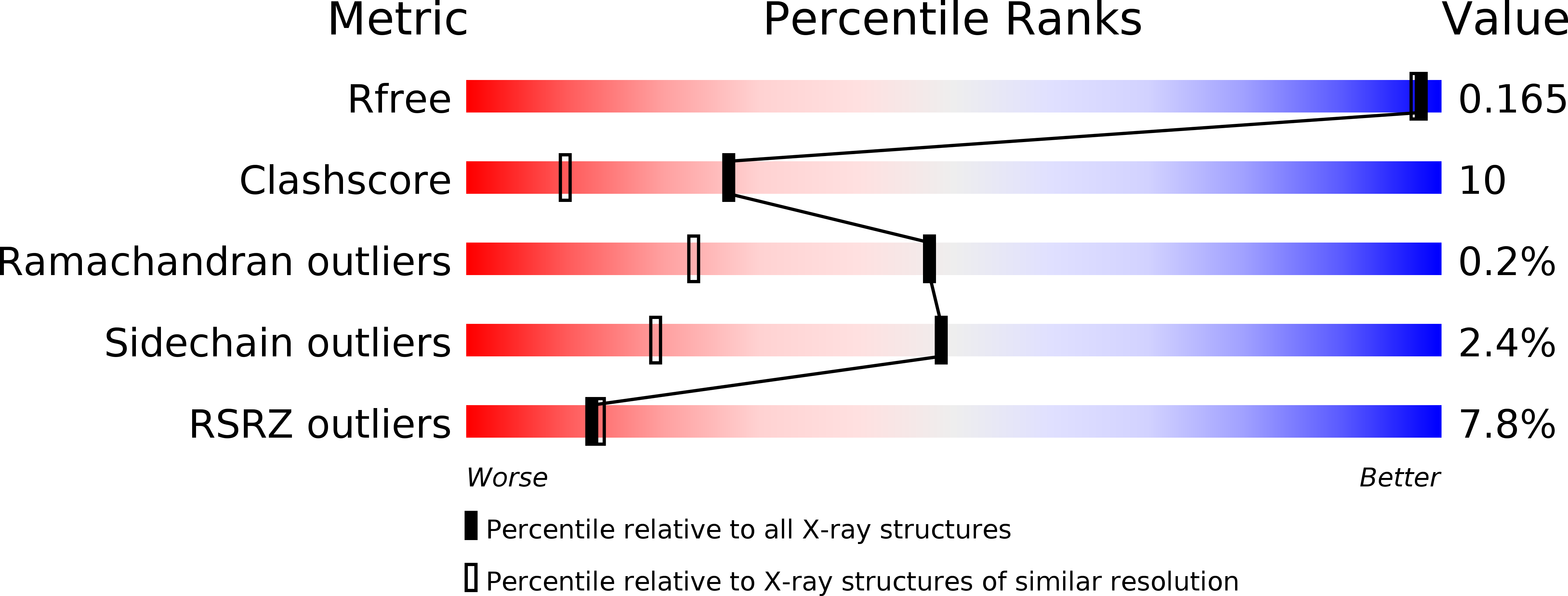
  • Method: X-RAY DIFFRACTION
  • Resolution: 1.50 Å
  • R-Value Free: 0.164 
  • R-Value Work: 0.113 
  • R-Value Observed: 0.116 

wwPDB Validation   3D Report Full Report


Ligand Structure Quality Assessment 


This is version 1.2 of the entry. See complete history


Literature

Novel Oxindole Sulfonamides and Sulfamides: EPZ031686, the First Orally Bioavailable Small Molecule SMYD3 Inhibitor.

Mitchell, L.H.Boriack-Sjodin, P.A.Smith, S.Thomenius, M.Rioux, N.Munchhof, M.Mills, J.E.Klaus, C.Totman, J.Riera, T.V.Raimondi, A.Jacques, S.L.West, K.Foley, M.Waters, N.J.Kuntz, K.W.Wigle, T.J.Scott, M.P.Copeland, R.A.Smith, J.J.Chesworth, R.

(2016) ACS Med Chem Lett 7: 134-138

  • DOI: https://doi.org/10.1021/acsmedchemlett.5b00272
  • Primary Citation of Related Structures:  
    5CCL, 5CCM

  • PubMed Abstract: 

    SMYD3 has been implicated in a range of cancers; however, until now no potent selective small molecule inhibitors have been available for target validation studies. A novel oxindole series of SMYD3 inhibitors was identified through screening of the Epizyme proprietary histone methyltransferase-biased library. Potency optimization afforded two tool compounds, sulfonamide EPZ031686 and sulfamide EPZ030456, with cellular potency at a level sufficient to probe the in vitro biology of SMYD3 inhibition. EPZ031686 shows good bioavailability following oral dosing in mice making it a suitable tool for potential in vivo target validation studies.


  • Organizational Affiliation

    Epizyme Inc. , Fourth Floor, 400 Technology Square, Cambridge, Massachusetts 02139, United States.


Macromolecules
Find similar proteins by:  (by identity cutoff)  |  3D Structure
Entity ID: 1
MoleculeChains Sequence LengthOrganismDetailsImage
Histone-lysine N-methyltransferase SMYD3432Homo sapiensMutation(s): 0 
Gene Names: SMYD3ZMYND1ZNFN3A1
EC: 2.1.1.43 (PDB Primary Data), 2.1.1.354 (UniProt)
UniProt & NIH Common Fund Data Resources
Find proteins for Q9H7B4 (Homo sapiens)
Explore Q9H7B4 
Go to UniProtKB:  Q9H7B4
PHAROS:  Q9H7B4
GTEx:  ENSG00000185420 
Entity Groups  
Sequence Clusters30% Identity50% Identity70% Identity90% Identity95% Identity100% Identity
UniProt GroupQ9H7B4
Sequence Annotations
Expand
  • Reference Sequence
Small Molecules
Ligands 4 Unique
IDChains Name / Formula / InChI Key2D Diagram3D Interactions
SAM
Query on SAM

Download Ideal Coordinates CCD File 
E [auth A]S-ADENOSYLMETHIONINE
C15 H22 N6 O5 S
MEFKEPWMEQBLKI-FCKMPRQPSA-N
4ZW
Query on 4ZW

Download Ideal Coordinates CCD File 
F [auth A],
G [auth A]
2-oxidanylidene-N-piperidin-4-yl-1,3-dihydroindole-5-carboxamide
C14 H17 N3 O2
SWMKOYKJOHHEQD-UHFFFAOYSA-N
ZN
Query on ZN

Download Ideal Coordinates CCD File 
B [auth A],
C [auth A],
D [auth A]
ZINC ION
Zn
PTFCDOFLOPIGGS-UHFFFAOYSA-N
EDO
Query on EDO

Download Ideal Coordinates CCD File 
H [auth A]1,2-ETHANEDIOL
C2 H6 O2
LYCAIKOWRPUZTN-UHFFFAOYSA-N
Binding Affinity Annotations 
IDSourceBinding Affinity
4ZW BindingDB:  5CCL IC50: min: 1.00e+4, max: 1.70e+4 (nM) from 3 assay(s)
Experimental Data & Validation

Experimental Data

  • Method: X-RAY DIFFRACTION
  • Resolution: 1.50 Å
  • R-Value Free: 0.164 
  • R-Value Work: 0.113 
  • R-Value Observed: 0.116 
  • Space Group: P 21 21 21
Unit Cell:
Length ( Å )Angle ( ˚ )
a = 58.594α = 90
b = 65.232β = 90
c = 105.966γ = 90
Software Package:
Software NamePurpose
HKL-2000data reduction
SCALEPACKdata scaling
REFMACrefinement
PDB_EXTRACTdata extraction

Structure Validation

View Full Validation Report


Ligand Structure Quality Assessment 


Entry History 

Deposition Data

Revision History  (Full details and data files)

  • Version 1.0: 2015-09-09
    Type: Initial release
  • Version 1.1: 2016-03-30
    Changes: Database references
  • Version 1.2: 2024-03-06
    Changes: Data collection, Database references, Derived calculations