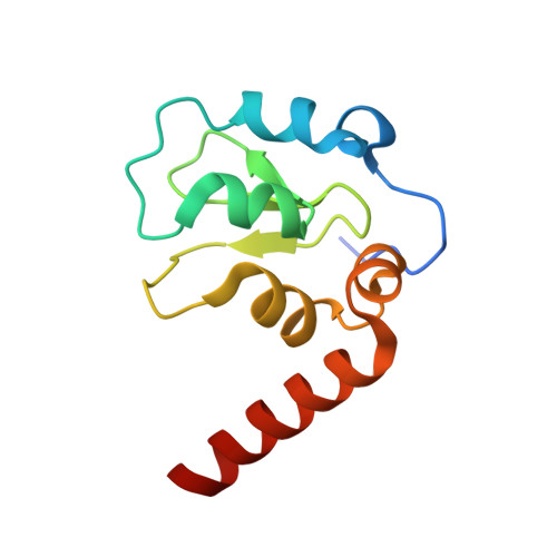
5C7D

Fragment-Based Drug Discovery Targeting Inhibitor of Apoptosis Proteins: Compound 17


Experimental Data Snapshot

  • Method: X-RAY DIFFRACTION
  • Resolution: 2.25 Å
  • R-Value Free: 0.251 
  • R-Value Work: 0.201 
  • R-Value Observed: 0.203 

wwPDB Validation   3D Report Full Report


Ligand Structure Quality Assessment 


This is version 1.2 of the entry. See complete history


Literature

Fragment-Based Drug Discovery Targeting Inhibitor of Apoptosis Proteins: Discovery of a Non-Alanine Lead Series with Dual Activity Against cIAP1 and XIAP.

Chessari, G.Buck, I.M.Day, J.E.Day, P.J.Iqbal, A.Johnson, C.N.Lewis, E.J.Martins, V.Miller, D.Reader, M.Rees, D.C.Rich, S.J.Tamanini, E.Vitorino, M.Ward, G.A.Williams, P.A.Williams, G.Wilsher, N.E.Woolford, A.J.

(2015) J Med Chem 58: 6574-6588

  • DOI: https://doi.org/10.1021/acs.jmedchem.5b00706
  • Primary Citation of Related Structures:  
    5C0K, 5C0L, 5C3H, 5C3K, 5C7A, 5C7B, 5C7C, 5C7D, 5C83, 5C84

  • PubMed Abstract: 

    Inhibitor of apoptosis proteins (IAPs) are important regulators of apoptosis and pro-survival signaling pathways whose deregulation is often associated with tumor genesis and tumor growth. IAPs have been proposed as targets for anticancer therapy, and a number of peptidomimetic IAP antagonists have entered clinical trials. Using our fragment-based screening approach, we identified nonpeptidic fragments binding with millimolar affinities to both cellular inhibitor of apoptosis protein 1 (cIAP1) and X-linked inhibitor of apoptosis protein (XIAP). Structure-based hit optimization together with an analysis of protein-ligand electrostatic potential complementarity allowed us to significantly increase binding affinity of the starting hits. Subsequent optimization gave a potent nonalanine IAP antagonist structurally distinct from all IAP antagonists previously reported. The lead compound had activity in cell-based assays and in a mouse xenograft efficacy model and represents a highly promising start point for further optimization.


  • Organizational Affiliation

    Astex Pharmaceuticals , 436 Cambridge Science Park, Milton Road, Cambridge, CB4 0QA, United Kingdom.


Macromolecules
Find similar proteins by:  (by identity cutoff)  |  3D Structure
Entity ID: 1
MoleculeChains Sequence LengthOrganismDetailsImage
E3 ubiquitin-protein ligase XIAP110Homo sapiensMutation(s): 0 
Gene Names: XIAPAPI3BIRC4IAP3
EC: 6.3.2 (PDB Primary Data), 2.3.2.27 (UniProt)
UniProt & NIH Common Fund Data Resources
Find proteins for P98170 (Homo sapiens)
Explore P98170 
Go to UniProtKB:  P98170
PHAROS:  P98170
GTEx:  ENSG00000101966 
Entity Groups  
Sequence Clusters30% Identity50% Identity70% Identity90% Identity95% Identity100% Identity
UniProt GroupP98170
Sequence Annotations
Expand
  • Reference Sequence
Small Molecules
Ligands 2 Unique
IDChains Name / Formula / InChI Key2D Diagram3D Interactions
4YF
Query on 4YF

Download Ideal Coordinates CCD File 
C [auth A](2R)-4-[2-(6-chloro-2,3-dihydro-1H-pyrrolo[3,2-c]pyridin-1-yl)-2-oxoethyl]-2-methylpiperazin-1-ium
C14 H20 Cl N4 O
UIUNSWTUDREESN-SNVBAGLBSA-O
ZN
Query on ZN

Download Ideal Coordinates CCD File 
B [auth A]ZINC ION
Zn
PTFCDOFLOPIGGS-UHFFFAOYSA-N
Experimental Data & Validation

Experimental Data

  • Method: X-RAY DIFFRACTION
  • Resolution: 2.25 Å
  • R-Value Free: 0.251 
  • R-Value Work: 0.201 
  • R-Value Observed: 0.203 
  • Space Group: P 41 2 2
Unit Cell:
Length ( Å )Angle ( ˚ )
a = 71.258α = 90
b = 71.258β = 90
c = 105.273γ = 90
Software Package:
Software NamePurpose
REFMACrefinement
d*TREKdata reduction
d*TREKdata scaling
PHASERphasing

Structure Validation

View Full Validation Report


Ligand Structure Quality Assessment 


Entry History 

Revision History  (Full details and data files)

  • Version 1.0: 2015-08-12
    Type: Initial release
  • Version 1.1: 2015-09-09
    Changes: Database references
  • Version 1.2: 2024-05-08
    Changes: Data collection, Database references