5BWN

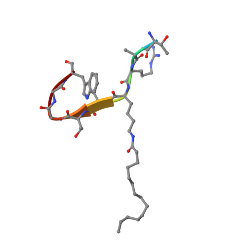
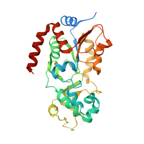
Crystal Structure of SIRT3 with a H3K9 Peptide Containing a Myristoyl Lysine


Experimental Data Snapshot

  • Method: X-RAY DIFFRACTION
  • Resolution: 1.94 Å
  • R-Value Free: 0.275 
  • R-Value Work: 0.226 
  • R-Value Observed: 0.231 

Starting Model: experimental
View more details

wwPDB Validation   3D Report Full Report


This is version 1.2 of the entry. See complete history


Literature

Crystal structures of SIRT3 reveal that the alpha 2-alpha 3 loop and alpha 3-helix affect the interaction with long-chain acyl lysine.

Gai, W.Li, H.Jiang, H.Long, Y.Liu, D.

(2016) FEBS Lett 590: 3019-3028

  • DOI: https://doi.org/10.1002/1873-3468.12345
  • Primary Citation of Related Structures:  
    5BWN, 5BWO

  • PubMed Abstract: 

    SIRT1-7 play important roles in many biological processes and age-related diseases. In addition to a NAD(+) -dependent deacetylase activity, they can catalyze several other reactions, including the hydrolysis of long-chain fatty acyl lysine. To study the binding modes of sirtuins to long-chain acyl lysines, we solved the crystal structures of SIRT3 bound to either a H3K9-myristoylated- or a H3K9-palmitoylated peptide. Interaction of SIRT3 with the palmitoyl group led to unfolding of the α3-helix. The myristoyl and palmitoyl groups bind to the C-pocket and an allosteric site near the α3-helix, respectively. We found that the residues preceding the α3-helix determine the size of the C-pocket. The flexibility of the α2-α3 loop and the plasticity of the α3-helix affect the interaction with long-chain acyl lysine.


  • Organizational Affiliation

    Department of Pharmacology III, Key Laboratory of Receptor Research, Shanghai Institute of Materia Medica, Chinese Academy of Sciences, China.


Macromolecules

Find similar proteins by:  Sequence   |   3D Structure  

Entity ID: 1
MoleculeChains Sequence LengthOrganismDetailsImage
myristoyl H3K9 peptideA [auth B]10synthetic constructMutation(s): 0 
UniProt & NIH Common Fund Data Resources
Find proteins for Q6NXT2 (Homo sapiens)
Explore Q6NXT2 
Go to UniProtKB:  Q6NXT2
PHAROS:  Q6NXT2
GTEx:  ENSG00000188375 
Entity Groups  
Sequence Clusters30% Identity50% Identity70% Identity90% Identity95% Identity100% Identity
UniProt GroupQ6NXT2
Sequence Annotations
Expand
  • Reference Sequence
Find similar proteins by:  (by identity cutoff)  |  3D Structure
Entity ID: 2
MoleculeChains Sequence LengthOrganismDetailsImage
NAD-dependent protein deacetylase sirtuin-3, mitochondrialB [auth A]309Homo sapiensMutation(s): 0 
Gene Names: SIRT3SIR2L3
EC: 3.5.1 (PDB Primary Data), 2.3.1.286 (UniProt)
UniProt & NIH Common Fund Data Resources
Find proteins for Q9NTG7 (Homo sapiens)
Explore Q9NTG7 
Go to UniProtKB:  Q9NTG7
PHAROS:  Q9NTG7
GTEx:  ENSG00000142082 
Entity Groups  
Sequence Clusters30% Identity50% Identity70% Identity90% Identity95% Identity100% Identity
UniProt GroupQ9NTG7
Sequence Annotations
Expand
  • Reference Sequence
Small Molecules
Ligands 1 Unique
IDChains Name / Formula / InChI Key2D Diagram3D Interactions
ZN
Query on ZN

Download Ideal Coordinates CCD File 
C [auth A]ZINC ION
Zn
PTFCDOFLOPIGGS-UHFFFAOYSA-N
Modified Residues  1 Unique
IDChains TypeFormula2D DiagramParent
MYK
Query on MYK
A [auth B]L-PEPTIDE LINKINGC20 H40 N2 O3LYS
Experimental Data & Validation

Experimental Data

  • Method: X-RAY DIFFRACTION
  • Resolution: 1.94 Å
  • R-Value Free: 0.275 
  • R-Value Work: 0.226 
  • R-Value Observed: 0.231 
  • Space Group: P 21 21 21
Unit Cell:
Length ( Å )Angle ( ˚ )
a = 44.102α = 90
b = 54.691β = 90
c = 117.257γ = 90
Software Package:
Software NamePurpose
PHENIXrefinement
HKL-2000data scaling
PHENIXmodel building

Structure Validation

View Full Validation Report


Entry History 

Deposition Data

Revision History  (Full details and data files)

  • Version 1.0: 2016-07-13
    Type: Initial release
  • Version 1.1: 2016-12-07
    Changes: Database references
  • Version 1.2: 2023-11-08
    Changes: Data collection, Database references, Refinement description