5B39

KIR3DL1*015 in complex with HLA-B*57:01


Experimental Data Snapshot

  • Method: X-RAY DIFFRACTION
  • Resolution: 2.50 Å
  • R-Value Free: 0.229 
  • R-Value Work: 0.178 
  • R-Value Observed: 0.181 

Starting Model: experimental
View more details

wwPDB Validation   3D Report Full Report


Ligand Structure Quality Assessment 


This is version 1.6 of the entry. See complete history


Literature

Killer cell immunoglobulin-like receptor 3DL1 polymorphism defines distinct hierarchies of HLA class I recognition

Saunders, P.M.Pymm, P.Pietra, G.Hughes, V.A.Hitchen, C.O'Connor, G.M.Loiacono, F.Widjaja, J.Price, D.A.Falco, M.Mingari, M.C.Moretta, L.McVicar, D.W.Rossjohn, J.Brooks, A.G.Vivian, J.P.

(2016) J Exp Med 213: 791-807

  • DOI: https://doi.org/10.1084/jem.20152023
  • Primary Citation of Related Structures:  
    5B38, 5B39

  • PubMed Abstract: 

    Natural killer (NK) cells play a key role in immunity, but how HLA class I (HLA-I) and killer cell immunoglobulin-like receptor 3DL1 (KIR3DL1) polymorphism impacts disease outcome remains unclear. KIR3DL1 (*001/*005/*015) tetramers were screened for reactivity against a panel of HLA-I molecules. This revealed different and distinct hierarchies of specificity for each KIR3DL1 allotype, with KIR3DL1*005 recognizing the widest array of HLA-I ligands. These differences were further reflected in functional studies using NK clones expressing these specific KIR3DL1 allotypes. Unexpectedly, the Ile/Thr80 dimorphism in the Bw4-motif did not categorically define strong/weak KIR3DL1 recognition. Although the KIR3DL1*001, *005, and *015 polymorphisms are remote from the KIR3DL1-HLA-I interface, the structures of these three KIR3DL1-HLA-I complexes showed that the broader HLA-I specificity of KIR3DL1*005 correlated with an altered KIR3DL1*005 interdomain positioning and increased mobility within its ligand-binding site. Collectively, we provide a generic framework for understanding the impact of KIR3DL1 polymorphism on the recognition of HLA-I allomorphs.


  • Organizational Affiliation

    Department of Microbiology and Immunology, Peter Doherty Institute for Infection and Immunity, The University of Melbourne, Parkville, Victoria 3010, Australia.


Macromolecules
Find similar proteins by:  (by identity cutoff)  |  3D Structure
Entity ID: 1
MoleculeChains Sequence LengthOrganismDetailsImage
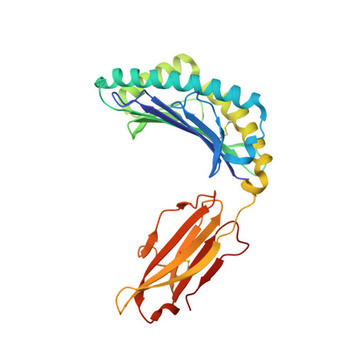
HLA class I histocompatibility antigen, B-57 alpha chain276Homo sapiensMutation(s): 0 
Gene Names: HLA-BHLAB
UniProt & NIH Common Fund Data Resources
Find proteins for P01889 (Homo sapiens)
Explore P01889 
Go to UniProtKB:  P01889
PHAROS:  P01889
GTEx:  ENSG00000234745 
Entity Groups  
Sequence Clusters30% Identity50% Identity70% Identity90% Identity95% Identity100% Identity
UniProt GroupP01889
Sequence Annotations
Expand
  • Reference Sequence
Find similar proteins by:  (by identity cutoff)  |  3D Structure
Entity ID: 2
MoleculeChains Sequence LengthOrganismDetailsImage
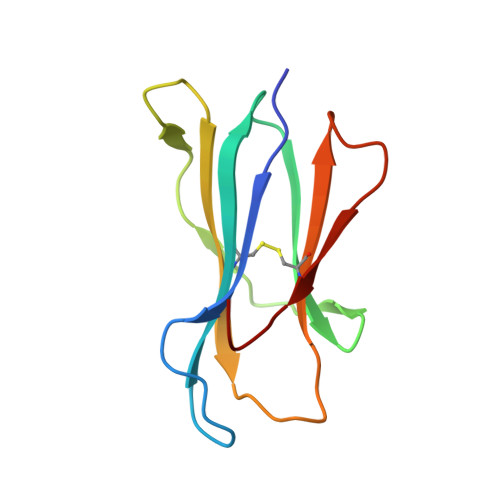
Beta-2-microglobulin99Homo sapiensMutation(s): 0 
Gene Names: B2MCDABP0092HDCMA22P
UniProt & NIH Common Fund Data Resources
Find proteins for P61769 (Homo sapiens)
Explore P61769 
Go to UniProtKB:  P61769
PHAROS:  P61769
GTEx:  ENSG00000166710 
Entity Groups  
Sequence Clusters30% Identity50% Identity70% Identity90% Identity95% Identity100% Identity
UniProt GroupP61769
Sequence Annotations
Expand
  • Reference Sequence

Find similar proteins by:  Sequence   |   3D Structure  

Entity ID: 3
MoleculeChains Sequence LengthOrganismDetailsImage
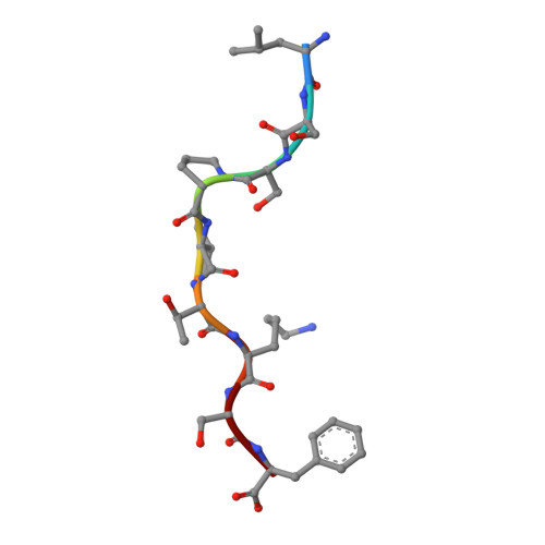
peptide from Ig kappa chain C region9Homo sapiensMutation(s): 0 
UniProt & NIH Common Fund Data Resources
Find proteins for P01834 (Homo sapiens)
Explore P01834 
Go to UniProtKB:  P01834
PHAROS:  P01834
Entity Groups  
UniProt GroupP01834
Sequence Annotations
Expand
  • Reference Sequence
Find similar proteins by:  (by identity cutoff)  |  3D Structure
Entity ID: 4
MoleculeChains Sequence LengthOrganismDetailsImage
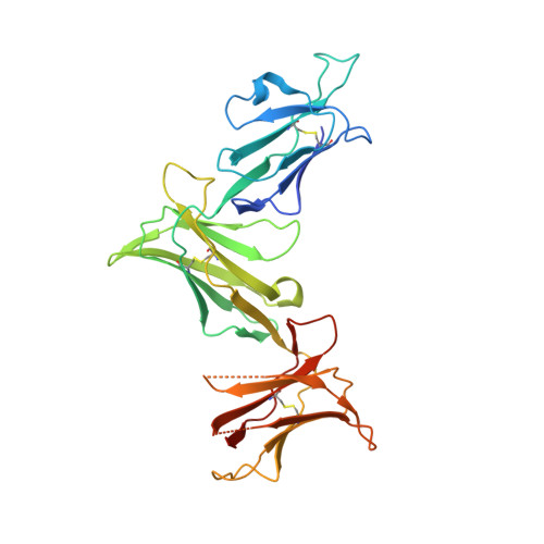
Killer cell immunoglobulin-like receptor 3DL1D [auth G]299Homo sapiensMutation(s): 3 
Gene Names: KIR3DL1CD158ENKAT3NKB1
UniProt & NIH Common Fund Data Resources
Find proteins for P43629 (Homo sapiens)
Explore P43629 
Go to UniProtKB:  P43629
PHAROS:  P43629
GTEx:  ENSG00000167633 
Entity Groups  
Sequence Clusters30% Identity50% Identity70% Identity90% Identity95% Identity100% Identity
UniProt GroupP43629
Glycosylation
Glycosylation Sites: 3Go to GlyGen: P43629-1
Sequence Annotations
Expand
  • Reference Sequence
Experimental Data & Validation

Experimental Data

  • Method: X-RAY DIFFRACTION
  • Resolution: 2.50 Å
  • R-Value Free: 0.229 
  • R-Value Work: 0.178 
  • R-Value Observed: 0.181 
  • Space Group: P 1
Unit Cell:
Length ( Å )Angle ( ˚ )
a = 51.568α = 95.56
b = 61.118β = 98.5
c = 65.089γ = 108.65
Software Package:
Software NamePurpose
PHENIXrefinement
MOSFLMdata reduction
SCALAdata scaling
PHASERphasing

Structure Validation

View Full Validation Report


Ligand Structure Quality Assessment 


Entry History & Funding Information

Deposition Data


Funding OrganizationLocationGrant Number
National Health and Medical Research Council (Australia)Australia1099814

Revision History  (Full details and data files)

  • Version 1.0: 2016-03-30
    Type: Initial release
  • Version 1.1: 2016-04-20
    Changes: Database references
  • Version 1.2: 2016-05-18
    Changes: Database references
  • Version 1.3: 2017-10-18
    Changes: Author supporting evidence, Data collection, Database references, Derived calculations
  • Version 1.4: 2020-07-29
    Type: Remediation
    Reason: Carbohydrate remediation
    Changes: Data collection, Derived calculations, Structure summary
  • Version 1.5: 2023-11-08
    Changes: Data collection, Database references, Refinement description, Structure summary
  • Version 1.6: 2024-11-06
    Changes: Structure summary