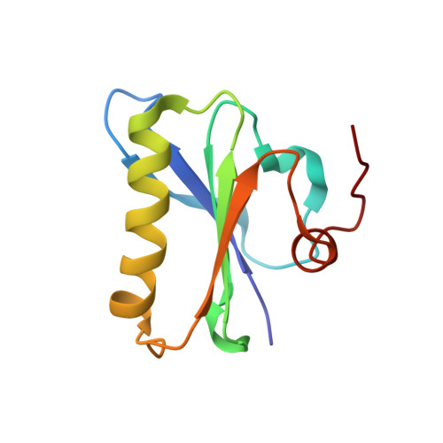
Calcium-Controlled Conformational Choreography in the N-Terminal Half of Adseverin.
Chumnarnsilpa, S., Robinson, R.C., Grimes, J.M., Leyrat, C.(2015) Nat Commun 6: 8254
- PubMed: 26365202
- DOI: https://doi.org/10.1038/ncomms9254
- Primary Citation of Related Structures:
5A1K, 5A1M - PubMed Abstract:
Adseverin is a member of the calcium-regulated gelsolin superfamily of actin-binding proteins. Here we report the crystal structure of the calcium-free N-terminal half of adseverin (iA1-A3) and the Ca(2+)-bound structure of A3, which reveal structural similarities and differences with gelsolin. Solution small-angle X-ray scattering combined with ensemble optimization revealed a dynamic Ca(2+)-dependent equilibrium between inactive, intermediate and active conformations. Increasing calcium concentrations progressively shift this equilibrium from a main population of inactive conformation to the active form. Molecular dynamics simulations of iA1-A3 provided insights into Ca(2+)-induced destabilization, implicating a critical role for the A2 type II calcium-binding site and the A2A3 linker in the activation process. Finally, mutations that disrupt the A1/A3 interface increase Ca(2+)-independent F-actin severing by A1-A3, albeit at a lower efficiency than observed for gelsolin domains G1-G3. Together, these data address the calcium dependency of A1-A3 activity in relation to the calcium-independent activity of G1-G3.
Organizational Affiliation:
Division of Structural Biology, University of Oxford, Henry Wellcome Building for Genomic Medicine, Oxford OX3 7BN, UK.