4ZQK

Structure of the complex of human programmed death-1 (PD-1) and its ligand PD-L1.


Experimental Data Snapshot

  • Method: X-RAY DIFFRACTION
  • Resolution: 2.45 Å
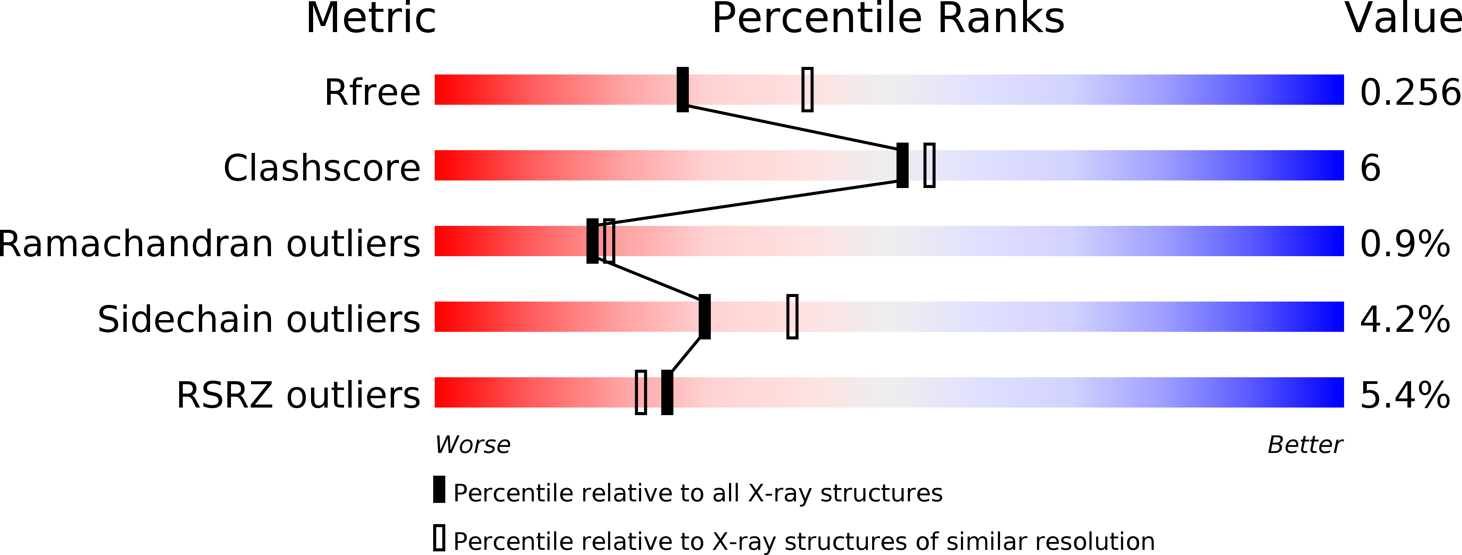
  • R-Value Free: 0.253 
  • R-Value Work: 0.207 
  • R-Value Observed: 0.209 

Starting Models: experimental
View more details

wwPDB Validation   3D Report Full Report


This is version 1.3 of the entry. See complete history


Literature

Structure of the Complex of Human Programmed Death 1, PD-1, and Its Ligand PD-L1.

Zak, K.M.Kitel, R.Przetocka, S.Golik, P.Guzik, K.Musielak, B.Domling, A.Dubin, G.Holak, T.A.

(2015) Structure 23: 2341-2348

  • DOI: https://doi.org/10.1016/j.str.2015.09.010
  • Primary Citation of Related Structures:  
    4ZQK, 5C3T

  • PubMed Abstract: 

    Targeting the PD-1/PD-L1 immunologic checkpoint with monoclonal antibodies has recently provided breakthrough progress in the treatment of melanoma, non-small cell lung cancer, and other types of cancer. Small-molecule drugs interfering with this pathway are highly awaited, but their development is hindered by insufficient structural information. This study reveals the molecular details of the human PD-1/PD-L1 interaction based on an X-ray structure of the complex. First, it is shown that the ligand binding to human PD-1 is associated with significant plasticity within the receptor. Second, a detailed molecular map of the interaction surface is provided, allowing definition of the regions within both interacting partners that may likely be targeted by small molecules.


  • Organizational Affiliation

    Faculty of Biochemistry, Biophysics and Biotechnology, Jagiellonian University, Gronostajowa 7, 30-387 Krakow, Poland; Malopolska Centre of Biotechnology, Jagiellonian University, Gronostajowa 7a, 30-387 Krakow, Poland.


Macromolecules
Find similar proteins by:  (by identity cutoff)  |  3D Structure
Entity ID: 1
MoleculeChains Sequence LengthOrganismDetailsImage
Programmed cell death 1 ligand 1115Homo sapiensMutation(s): 0 
Gene Names: CD274B7H1PDCD1L1PDCD1LG1PDL1
UniProt & NIH Common Fund Data Resources
Find proteins for Q9NZQ7 (Homo sapiens)
Explore Q9NZQ7 
Go to UniProtKB:  Q9NZQ7
GTEx:  ENSG00000120217 
Entity Groups  
Sequence Clusters30% Identity50% Identity70% Identity90% Identity95% Identity100% Identity
UniProt GroupQ9NZQ7
Sequence Annotations
Expand
  • Reference Sequence
Find similar proteins by:  (by identity cutoff)  |  3D Structure
Entity ID: 2
MoleculeChains Sequence LengthOrganismDetailsImage
Programmed cell death protein 1118Homo sapiensMutation(s): 1 
Gene Names: PDCD1PD1
UniProt & NIH Common Fund Data Resources
Find proteins for Q15116 (Homo sapiens)
Explore Q15116 
Go to UniProtKB:  Q15116
GTEx:  ENSG00000188389 
Entity Groups  
Sequence Clusters30% Identity50% Identity70% Identity90% Identity95% Identity100% Identity
UniProt GroupQ15116
Sequence Annotations
Expand
  • Reference Sequence
Small Molecules
Ligands 1 Unique
IDChains Name / Formula / InChI Key2D Diagram3D Interactions
NA
Query on NA

Download Ideal Coordinates CCD File 
C [auth A]SODIUM ION
Na
FKNQFGJONOIPTF-UHFFFAOYSA-N
Experimental Data & Validation

Experimental Data

  • Method: X-RAY DIFFRACTION
  • Resolution: 2.45 Å
  • R-Value Free: 0.253 
  • R-Value Work: 0.207 
  • R-Value Observed: 0.209 
  • Space Group: P 31 2 1
Unit Cell:
Length ( Å )Angle ( ˚ )
a = 70.862α = 90
b = 70.862β = 90
c = 114.367γ = 120
Software Package:
Software NamePurpose
Aimlessdata scaling
PHASERphasing
REFMACrefinement
PDB_EXTRACTdata extraction
XDSdata reduction

Structure Validation

View Full Validation Report


Entry History 

Deposition Data

Revision History  (Full details and data files)

  • Version 1.0: 2015-11-04
    Type: Initial release
  • Version 1.1: 2015-12-09
    Changes: Database references
  • Version 1.2: 2015-12-16
    Changes: Database references
  • Version 1.3: 2024-01-10
    Changes: Data collection, Database references, Refinement description