4ZMT

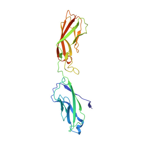
Crystal structure of human P-cadherin (ss-X-dimer-long)


Experimental Data Snapshot

  • Method: X-RAY DIFFRACTION
  • Resolution: 2.70 Å
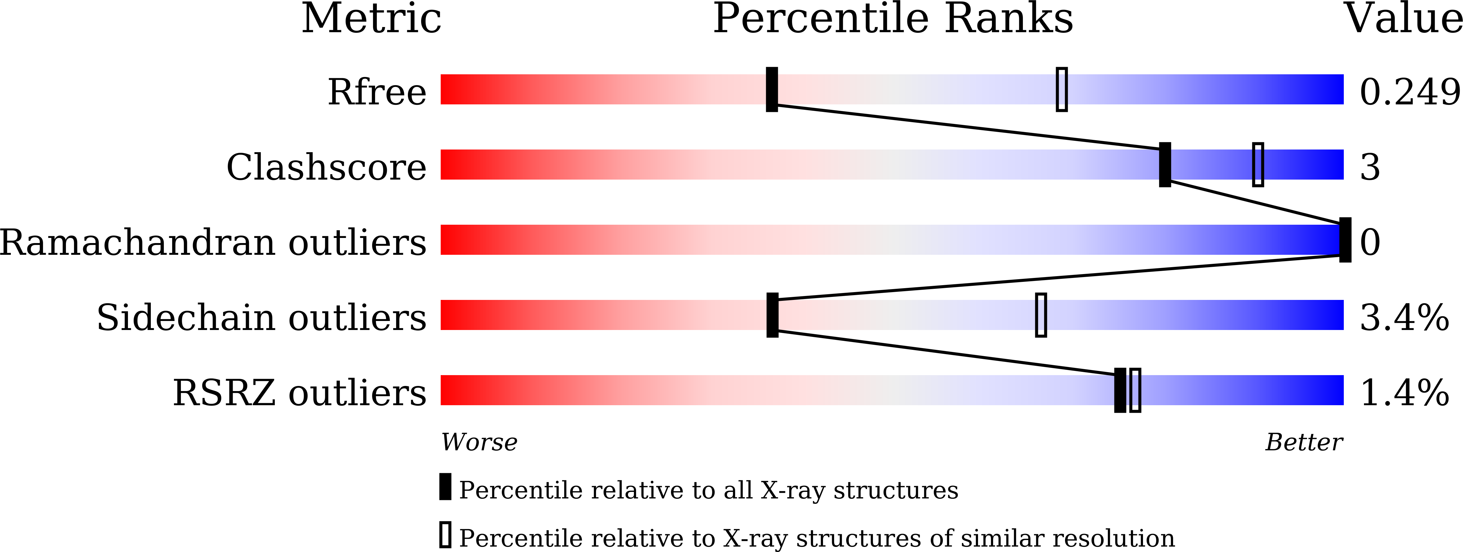
  • R-Value Free: 0.249 
  • R-Value Work: 0.228 
  • R-Value Observed: 0.229 

Starting Model: experimental
View more details

wwPDB Validation   3D Report Full Report


This is version 1.3 of the entry. See complete history


Literature

Adhesive Dimerization of Human P-Cadherin Catalyzed by a Chaperone-like Mechanism

Kudo, S.Caaveiro, J.M.Tsumoto, K.

(2016) Structure 24: 1523-1536

  • DOI: https://doi.org/10.1016/j.str.2016.07.002
  • Primary Citation of Related Structures:  
    4ZML, 4ZMN, 4ZMO, 4ZMP, 4ZMQ, 4ZMT, 4ZMV, 4ZMW, 4ZMX, 4ZMY, 4ZMZ

  • PubMed Abstract: 

    Orderly assembly of classical cadherins governs cell adhesion and tissue maintenance. A key event is the strand-swap dimerization of the extracellular ectodomains of two cadherin molecules from apposing cells. Here we have determined crystal structures of P-cadherin in six different conformational states to elaborate a motion picture of its adhesive dimerization at the atomic level. The snapshots revealed that cell-adhesive dimerization is facilitated by several intermediate states collectively termed X-dimer in analogy to other classical cadherins. Based on previous studies and on the combined structural, kinetic, thermodynamic, biochemical, and cellular data reported herein, we propose that the adhesive dimerization of human P-cadherin is achieved by a stepwise mechanism analogous to that of assembly chaperones. This mechanism, applicable to type I classical cadherins, confers high specificity and fast association rates. We expect these findings to guide innovative therapeutic approaches targeting P-cadherin in cancer.


  • Organizational Affiliation

    Department of Chemistry & Biotechnology, The University of Tokyo, Tokyo 108-8639, Japan.


Macromolecules
Find similar proteins by:  (by identity cutoff)  |  3D Structure
Entity ID: 1
MoleculeChains Sequence LengthOrganismDetailsImage
Cadherin-3
A, B, C, D, E
A, B, C, D, E, F
232Homo sapiensMutation(s): 0 
Gene Names: CDH3CDHP
UniProt & NIH Common Fund Data Resources
Find proteins for P22223 (Homo sapiens)
Explore P22223 
Go to UniProtKB:  P22223
PHAROS:  P22223
GTEx:  ENSG00000062038 
Entity Groups  
Sequence Clusters30% Identity50% Identity70% Identity90% Identity95% Identity100% Identity
UniProt GroupP22223
Sequence Annotations
Expand
  • Reference Sequence
Small Molecules
Ligands 1 Unique
IDChains Name / Formula / InChI Key2D Diagram3D Interactions
CA
Query on CA

Download Ideal Coordinates CCD File 
G [auth A]
H [auth A]
I [auth A]
J [auth B]
K [auth B]
G [auth A],
H [auth A],
I [auth A],
J [auth B],
K [auth B],
L [auth B],
M [auth C],
N [auth C],
O [auth C],
P [auth D],
Q [auth D],
R [auth D],
S [auth E],
T [auth E],
U [auth E],
V [auth F],
W [auth F],
X [auth F]
CALCIUM ION
Ca
BHPQYMZQTOCNFJ-UHFFFAOYSA-N
Experimental Data & Validation

Experimental Data

  • Method: X-RAY DIFFRACTION
  • Resolution: 2.70 Å
  • R-Value Free: 0.249 
  • R-Value Work: 0.228 
  • R-Value Observed: 0.229 
  • Space Group: P 1 21 1
Unit Cell:
Length ( Å )Angle ( ˚ )
a = 85.21α = 90
b = 105.83β = 96.8
c = 90.21γ = 90
Software Package:
Software NamePurpose
REFMACrefinement
MOSFLMdata reduction
SCALAdata scaling
PHASERphasing

Structure Validation

View Full Validation Report


Entry History 

Deposition Data

Revision History  (Full details and data files)

  • Version 1.0: 2016-09-07
    Type: Initial release
  • Version 1.1: 2016-09-21
    Changes: Database references
  • Version 1.2: 2020-02-19
    Changes: Data collection, Database references, Derived calculations
  • Version 1.3: 2023-11-08
    Changes: Data collection, Database references, Derived calculations, Refinement description