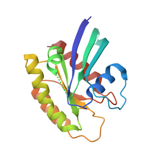
Tyrosine phosphorylation of RAS by ABL allosterically enhances effector binding.
Ting, P.Y., Johnson, C.W., Fang, C., Cao, X., Graeber, T.G., Mattos, C., Colicelli, J.(2015) FASEB J 29: 3750-3761
- PubMed: 25999467
- DOI: https://doi.org/10.1096/fj.15-271510
- Primary Citation of Related Structures:
4XVQ, 4XVR - PubMed Abstract:
RAS proteins are signal transduction gatekeepers that mediate cell growth, survival, and differentiation through interactions with multiple effector proteins. The RAS effector RAS- and RAB-interacting protein 1 (RIN1) activates its own downstream effectors, the small GTPase RAB5 and the tyrosine kinase Abelson tyrosine-protein kinase (ABL), to modulate endocytosis and cytoskeleton remodeling. To identify ABL substrates downstream of RAS-to-RIN1 signaling, we examined human HEK293T cells overexpressing components of this pathway. Proteomic analysis revealed several novel phosphotyrosine peptides, including Harvey rat sarcoma oncogene (HRAS)-pTyr(137). Here we report that ABL phosphorylates tyrosine 137 of H-, K-, and NRAS. Increased RIN1 levels enhanced HRAS-Tyr(137) phosphorylation by nearly 5-fold, suggesting that RAS-stimulated RIN1 can drive ABL-mediated RAS modification in a feedback circuit. Tyr(137) is well conserved among RAS orthologs and is part of a transprotein H-bond network. Crystal structures of HRAS(Y137F) and HRAS(Y137E) revealed conformation changes radiating from the mutated residue. Although consistent with Tyr(137) participation in allosteric control of HRAS function, the mutations did not alter intrinsic GTP hydrolysis rates in vitro. HRAS-Tyr(137) phosphorylation enhanced HRAS signaling capacity in cells, however, as reflected by a 4-fold increase in the association of phosphorylated HRAS(G12V) with its effector protein RAF proto-oncogene serine/threonine protein kinase 1 (RAF1). These data suggest that RAS phosphorylation at Tyr(137) allosterically alters protein conformation and effector binding, providing a mechanism for effector-initiated modulation of RAS signaling.
Organizational Affiliation:
*Molecular Biology Institute, Jonsson Comprehensive Cancer Center, Department of Biological Chemistry, David Geffen School of Medicine, University of California, Los Angeles, Los Angeles, California, USA; Department of Chemistry and Chemical Biology, Northeastern University, Boston, Massachusetts, USA; and Department of Molecular and Medical Pharmacology, Crump Institute for Molecular Imaging, University of California at Los Angeles Metabolomics and Proteomics Center, California Nanosystems Institute and Jonsson Comprehensive Cancer Center, University of California, Los Angeles, Los Angeles, California, USA.


