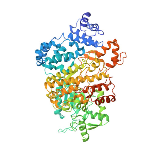
4X3V

Crystal structure of human ribonucleotide reductase 1 bound to inhibitor


Experimental Data Snapshot

  • Method: X-RAY DIFFRACTION
  • Resolution: 3.70 Å
  • R-Value Free: 0.289 
  • R-Value Work: 0.232 
  • R-Value Observed: 0.238 

Starting Model: experimental
View more details

wwPDB Validation   3D Report Full Report


Ligand Structure Quality Assessment 


This is version 1.4 of the entry. See complete history


Literature

Identification of Non-nucleoside Human Ribonucleotide Reductase Modulators.

Ahmad, M.F.Huff, S.E.Pink, J.Alam, I.Zhang, A.Perry, K.Harris, M.E.Misko, T.Porwal, S.K.Oleinick, N.L.Miyagi, M.Viswanathan, R.Dealwis, C.G.

(2015) J Med Chem 58: 9498-9509

  • DOI: https://doi.org/10.1021/acs.jmedchem.5b00929
  • Primary Citation of Related Structures:  
    4X3V

  • PubMed Abstract: 

    Ribonucleotide reductase (RR) catalyzes the rate-limiting step of dNTP synthesis and is an established cancer target. Drugs targeting RR are mainly nucleoside in nature. In this study, we sought to identify non-nucleoside small-molecule inhibitors of RR. Using virtual screening, binding affinity, inhibition, and cell toxicity, we have discovered a class of small molecules that alter the equilibrium of inactive hexamers of RR, leading to its inhibition. Several unique chemical categories, including a phthalimide derivative, show micromolar IC50s and KDs while demonstrating cytotoxicity. A crystal structure of an active phthalimide binding at the targeted interface supports the noncompetitive mode of inhibition determined by kinetic studies. Furthermore, the phthalimide shifts the equilibrium from dimer to hexamer. Together, these data identify several novel non-nucleoside inhibitors of human RR which act by stabilizing the inactive form of the enzyme.


  • Organizational Affiliation

    Department of Pharmacology, School of Medicine, Case Western Reserve University , Cleveland, Ohio 44106, United States.


Macromolecules
Find similar proteins by:  (by identity cutoff)  |  3D Structure
Entity ID: 1
MoleculeChains Sequence LengthOrganismDetailsImage
Ribonucleoside-diphosphate reductase large subunit
A, B
792Homo sapiensMutation(s): 0 
Gene Names: RRM1RR1
EC: 1.17.4.1
UniProt & NIH Common Fund Data Resources
Find proteins for P23921 (Homo sapiens)
Go to UniProtKB:  P23921
PHAROS:  P23921
GTEx:  ENSG00000167325 
Sequence Annotations
Expand
  • Reference Sequence
Small Molecules
Ligands 2 Unique
IDChains Name / Formula / InChI Key2D Diagram3D Interactions
TTP
Query on TTP

Download Ideal Coordinates CCD File 
C [auth A],
E [auth B]
THYMIDINE-5'-TRIPHOSPHATE
C10 H17 N2 O14 P3
NHVNXKFIZYSCEB-XLPZGREQSA-N
3X4
Query on 3X4

Download Ideal Coordinates CCD File 
D [auth A]N~6~-{N-[(1,3-dioxo-1,3-dihydro-2H-isoindol-2-yl)acetyl]-2-methyl-D-alanyl}-D-lysine
C20 H26 N4 O6
HSKHXKLBWCBTSW-CQSZACIVSA-N
Experimental Data & Validation

Experimental Data

  • Method: X-RAY DIFFRACTION
  • Resolution: 3.70 Å
  • R-Value Free: 0.289 
  • R-Value Work: 0.232 
  • R-Value Observed: 0.238 
  • Space Group: P 21 21 21
Unit Cell:
Length ( Å )Angle ( ˚ )
a = 71.88α = 90
b = 110.498β = 90
c = 216.721γ = 90
Software Package:
Software NamePurpose
PHENIXrefinement
REFMACphasing

Structure Validation

View Full Validation Report


Ligand Structure Quality Assessment 


Entry History & Funding Information

Deposition Data


Funding OrganizationLocationGrant Number
National Institutes of Health/National Institute of General Medical Sciences (NIH/NIGMS)United StatesR01GM100887-01
National Institutes of Health/National Cancer Institute (NIH/NCI)United StatesR01CA100827-04A1

Revision History  (Full details and data files)

  • Version 1.0: 2015-11-11
    Type: Initial release
  • Version 1.1: 2016-01-06
    Changes: Database references
  • Version 1.2: 2017-09-13
    Changes: Advisory, Author supporting evidence, Database references, Derived calculations
  • Version 1.3: 2019-12-04
    Changes: Author supporting evidence
  • Version 1.4: 2023-09-27
    Changes: Advisory, Data collection, Database references, Refinement description