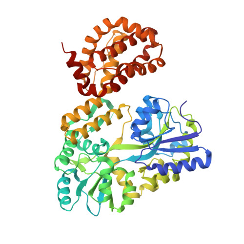
4WMV

STRUCTURE OF MBP-MCL1 BOUND TO ligand 4 AT 2.4A


Experimental Data Snapshot

  • Method: X-RAY DIFFRACTION
  • Resolution: 2.40 Å
  • R-Value Free: 0.253 
  • R-Value Work: 0.178 
  • R-Value Observed: 0.182 

Starting Models: experimental
View more details

wwPDB Validation   3D Report Full Report


Ligand Structure Quality Assessment 


This is version 2.1 of the entry. See complete history


Literature

A Maltose-Binding Protein Fusion Construct Yields a Robust Crystallography Platform for MCL1.

Clifton, M.C.Dranow, D.M.Leed, A.Fulroth, B.Fairman, J.W.Abendroth, J.Atkins, K.A.Wallace, E.Fan, D.Xu, G.Ni, Z.J.Daniels, D.Van Drie, J.Wei, G.Burgin, A.B.Golub, T.R.Hubbard, B.K.Serrano-Wu, M.H.

(2015) PLoS One 10: e0125010-e0125010

  • DOI: https://doi.org/10.1371/journal.pone.0125010
  • Primary Citation of Related Structures:  
    4WMR, 4WMS, 4WMT, 4WMU, 4WMV, 4WMW, 4WMX

  • PubMed Abstract: 

    Crystallization of a maltose-binding protein MCL1 fusion has yielded a robust crystallography platform that generated the first apo MCL1 crystal structure, as well as five ligand-bound structures. The ability to obtain fragment-bound structures advances structure-based drug design efforts that, despite considerable effort, had previously been intractable by crystallography. In the ligand-independent crystal form we identify inhibitor binding modes not observed in earlier crystallographic systems. This MBP-MCL1 construct dramatically improves the structural understanding of well-validated MCL1 ligands, and will likely catalyze the structure-based optimization of high affinity MCL1 inhibitors.


  • Organizational Affiliation

    Beryllium, Bedford, Massachusetts, United States of America.


Macromolecules
Find similar proteins by:  (by identity cutoff)  |  3D Structure
Entity ID: 1
MoleculeChains Sequence LengthOrganismDetailsImage
MBL-MCL1 chimera protein518Escherichia coli K-12Homo sapiens
This entity is chimeric
Mutation(s): 3 
Gene Names: malEb4034JW3994MCL1BCL2L3
UniProt & NIH Common Fund Data Resources
Find proteins for P0AEX9 (Escherichia coli (strain K12))
Explore P0AEX9 
Go to UniProtKB:  P0AEX9
Find proteins for Q07820 (Homo sapiens)
Explore Q07820 
Go to UniProtKB:  Q07820
PHAROS:  Q07820
GTEx:  ENSG00000143384 
Entity Groups  
Sequence Clusters30% Identity50% Identity70% Identity90% Identity95% Identity100% Identity
UniProt GroupsP0AEX9Q07820
Sequence Annotations
Expand
  • Reference Sequence
Oligosaccharides

Help

Entity ID: 2
MoleculeChains Length2D Diagram Glycosylation3D Interactions
alpha-D-glucopyranose-(1-4)-alpha-D-glucopyranose
B
2N/A
Glycosylation Resources
GlyTouCan:  G07411ON
GlyCosmos:  G07411ON
Small Molecules
Ligands 3 Unique
IDChains Name / Formula / InChI Key2D Diagram3D Interactions
3R4
Query on 3R4

Download Ideal Coordinates CCD File 
E [auth A]3-chloro-6-fluoro-1-benzothiophene-2-carboxylic acid
C9 H4 Cl F O2 S
HSRSWUJIPYUCSE-UHFFFAOYSA-N
CL
Query on CL

Download Ideal Coordinates CCD File 
C [auth A]CHLORIDE ION
Cl
VEXZGXHMUGYJMC-UHFFFAOYSA-M
MG
Query on MG

Download Ideal Coordinates CCD File 
D [auth A]MAGNESIUM ION
Mg
JLVVSXFLKOJNIY-UHFFFAOYSA-N
Biologically Interesting Molecules (External Reference) 1 Unique
Entity ID: 2
IDChains NameType/Class2D Diagram3D Interactions
PRD_900001
Query on PRD_900001
B
alpha-maltoseOligosaccharide / Nutrient
Experimental Data & Validation

Experimental Data

  • Method: X-RAY DIFFRACTION
  • Resolution: 2.40 Å
  • R-Value Free: 0.253 
  • R-Value Work: 0.178 
  • R-Value Observed: 0.182 
  • Space Group: P 21 21 2
Unit Cell:
Length ( Å )Angle ( ˚ )
a = 98.89α = 90
b = 136.07β = 90
c = 37.82γ = 90
Software Package:
Software NamePurpose
PHENIXrefinement
XDSdata reduction
PDB_EXTRACTdata extraction

Structure Validation

View Full Validation Report


Ligand Structure Quality Assessment 


Entry History 

Deposition Data

Revision History  (Full details and data files)

  • Version 1.0: 2015-05-06
    Type: Initial release
  • Version 2.0: 2020-07-29
    Type: Remediation
    Reason: Carbohydrate remediation
    Changes: Atomic model, Data collection, Derived calculations, Non-polymer description, Other, Refinement description, Source and taxonomy, Structure summary
  • Version 2.1: 2023-09-27
    Changes: Data collection, Database references, Refinement description, Structure summary