4RG9

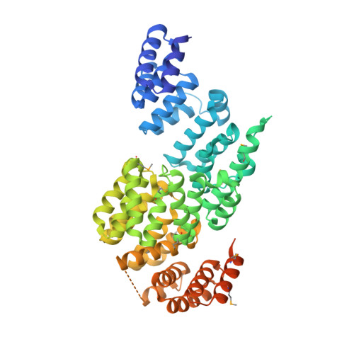
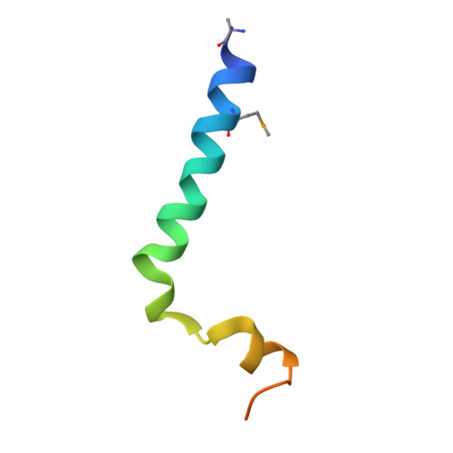
Crystal structure of APC3-APC16 complex (Selenomethionine Derivative)


Experimental Data Snapshot

  • Method: X-RAY DIFFRACTION
  • Resolution: 3.25 Å
  • R-Value Free: 0.248 
  • R-Value Work: 0.210 
  • R-Value Observed: 0.212 

wwPDB Validation   3D Report Full Report


This is version 1.3 of the entry. See complete history


Literature

Structure of an APC3-APC16 Complex: Insights into Assembly of the Anaphase-Promoting Complex/Cyclosome.

Yamaguchi, M.Yu, S.Qiao, R.Weissmann, F.Miller, D.J.VanderLinden, R.Brown, N.G.Frye, J.J.Peters, J.M.Schulman, B.A.

(2015) J Mol Biol 427: 1748-1764

  • DOI: https://doi.org/10.1016/j.jmb.2014.11.020
  • Primary Citation of Related Structures:  
    4RG6, 4RG7, 4RG9

  • PubMed Abstract: 

    The anaphase-promoting complex/cyclosome (APC/C) is a massive E3 ligase that controls mitosis by catalyzing ubiquitination of key cell cycle regulatory proteins. The APC/C assembly contains two subcomplexes: the "Platform" centers around a cullin-RING-like E3 ligase catalytic core; the "Arc Lamp" is a hub that mediates transient association with regulators and ubiquitination substrates. The Arc Lamp contains the small subunits APC16, CDC26, and APC13, and tetratricopeptide repeat (TPR) proteins (APC7, APC3, APC6, and APC8) that homodimerize and stack with quasi-2-fold symmetry. Within the APC/C complex, APC3 serves as center for regulation. APC3's TPR motifs recruit substrate-binding coactivators, CDC20 and CDH1, via their C-terminal conserved Ile-Arg (IR) tail sequences. Human APC3 also binds APC16 and APC7 and contains a >200-residue loop that is heavily phosphorylated during mitosis, although the basis for APC3 interactions and whether loop phosphorylation is required for ubiquitination are unclear. Here, we map the basis for human APC3 assembly with APC16 and APC7, report crystal structures of APC3Δloop alone and in complex with the C-terminal domain of APC16, and test roles of APC3's loop and IR tail binding surfaces in APC/C-catalyzed ubiquitination. The structures show how one APC16 binds asymmetrically to the symmetric APC3 dimer and, together with biochemistry and prior data, explain how APC16 recruits APC7 to APC3, show how APC3's C-terminal domain is rearranged in the full APC/C assembly, and visualize residues in the IR tail binding cleft important for coactivator-dependent ubiquitination. Overall, the results provide insights into assembly, regulation, and interactions of TPR proteins and the APC/C.


  • Organizational Affiliation

    Department of Structural Biology, St. Jude Children's Research Hospital, Memphis, TN 38105, USA.


Macromolecules
Find similar proteins by:  (by identity cutoff)  |  3D Structure
Entity ID: 1
MoleculeChains Sequence LengthOrganismDetailsImage
Cell division cycle protein 27 homolog
A, B
560Homo sapiensMutation(s): 0 
Gene Names: CDC27ANAPC3D0S1430ED17S978E
UniProt & NIH Common Fund Data Resources
Find proteins for P30260 (Homo sapiens)
Explore P30260 
Go to UniProtKB:  P30260
PHAROS:  P30260
GTEx:  ENSG00000004897 
Entity Groups  
Sequence Clusters30% Identity50% Identity70% Identity90% Identity95% Identity100% Identity
UniProt GroupP30260
Sequence Annotations
Expand
  • Reference Sequence
Find similar proteins by:  (by identity cutoff)  |  3D Structure
Entity ID: 2
MoleculeChains Sequence LengthOrganismDetailsImage
Anaphase-promoting complex subunit 16C [auth S]43Homo sapiensMutation(s): 0 
Gene Names: ANAPC16C10orf104
UniProt & NIH Common Fund Data Resources
Find proteins for Q96DE5 (Homo sapiens)
Explore Q96DE5 
Go to UniProtKB:  Q96DE5
PHAROS:  Q96DE5
GTEx:  ENSG00000166295 
Entity Groups  
Sequence Clusters30% Identity50% Identity70% Identity90% Identity95% Identity100% Identity
UniProt GroupQ96DE5
Sequence Annotations
Expand
  • Reference Sequence
Small Molecules
Modified Residues  1 Unique
IDChains TypeFormula2D DiagramParent
MSE
Query on MSE
A, B
L-PEPTIDE LINKINGC5 H11 N O2 SeMET
Experimental Data & Validation

Experimental Data

  • Method: X-RAY DIFFRACTION
  • Resolution: 3.25 Å
  • R-Value Free: 0.248 
  • R-Value Work: 0.210 
  • R-Value Observed: 0.212 
  • Space Group: P 43
Unit Cell:
Length ( Å )Angle ( ˚ )
a = 116.019α = 90
b = 116.019β = 90
c = 184.352γ = 90
Software Package:
Software NamePurpose
XDSdata scaling
SHELXmodel building
REFMACrefinement
pointlessdata scaling
SCALAdata scaling
SHELXphasing

Structure Validation

View Full Validation Report


Entry History 

Deposition Data

Revision History  (Full details and data files)

  • Version 1.0: 2014-12-24
    Type: Initial release
  • Version 1.1: 2015-01-14
    Changes: Database references
  • Version 1.2: 2015-04-15
    Changes: Database references
  • Version 1.3: 2017-08-02
    Changes: Refinement description, Source and taxonomy