4QMG

The Structure of MTDH-SND1 Complex Reveals Novel Cancer-Promoting Interactions


Experimental Data Snapshot

  • Method: X-RAY DIFFRACTION
  • Resolution: 2.70 Å
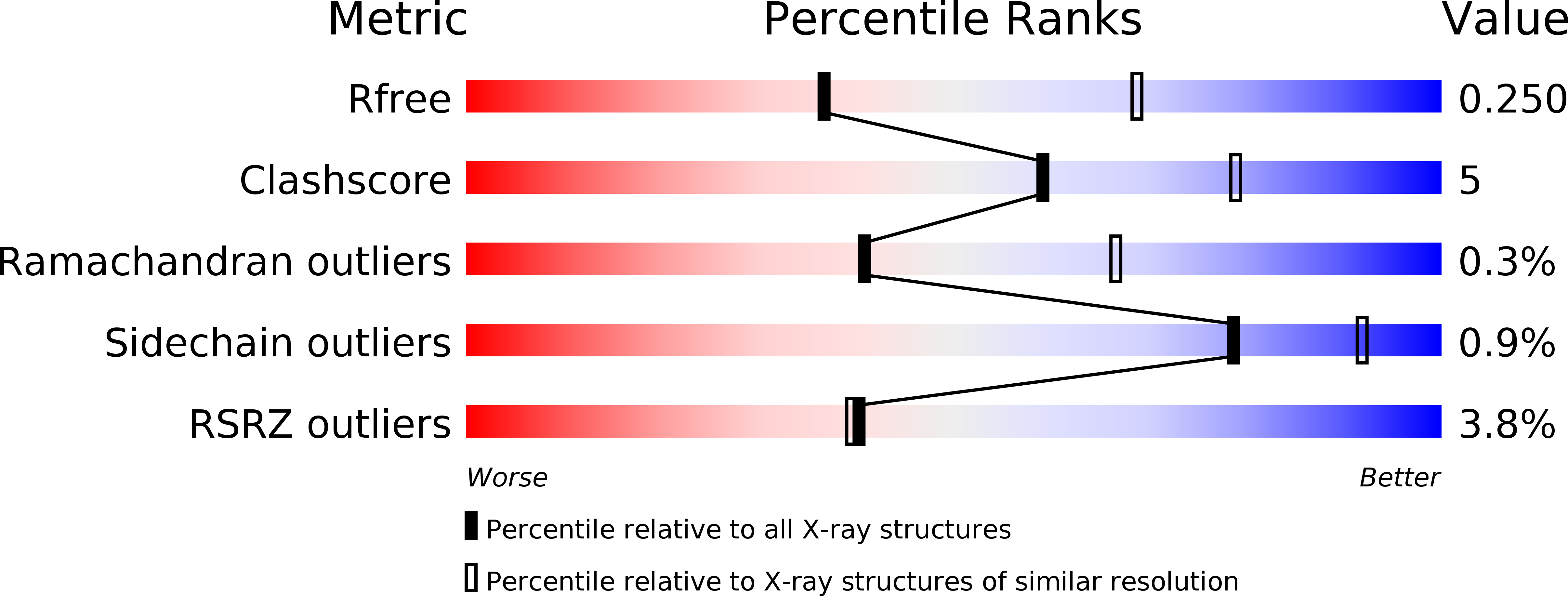
  • R-Value Free: 0.248 
  • R-Value Work: 0.190 
  • R-Value Observed: 0.192 

wwPDB Validation   3D Report Full Report


This is version 1.0 of the entry. See complete history


Literature

Structural Insights into the Tumor-Promoting Function of the MTDH-SND1 Complex.

Guo, F.Wan, L.Zheng, A.Stanevich, V.Wei, Y.Satyshur, K.A.Shen, M.Lee, W.Kang, Y.Xing, Y.

(2014) Cell Rep 8: 1704-1713

  • DOI: https://doi.org/10.1016/j.celrep.2014.08.033
  • Primary Citation of Related Structures:  
    4QMG

  • PubMed Abstract: 

    Metadherin (MTDH) and Staphylococcal nuclease domain containing 1 (SND1) are overexpressed and interact in diverse cancer types. The structural mechanism of their interaction remains unclear. Here, we determined the high-resolution crystal structure of MTDH-SND1 complex, which reveals an 11-residue MTDH peptide motif occupying an extended protein groove between two SN domains (SN1/2), with two MTDH tryptophan residues nestled into two well-defined pockets in SND1. At the opposite side of the MTDH-SND1 binding interface, SND1 possesses long protruding arms and deep surface valleys that are prone to binding with other partners. Despite the simple binding mode, interactions at both tryptophan-binding pockets are important for MTDH and SND1's roles in breast cancer and for SND1 stability under stress. Our study reveals a unique mode of interaction with SN domains that dictates cancer-promoting activity and provides a structural basis for mechanistic understanding of MTDH-SND1-mediated signaling and for exploring therapeutic targeting of this complex.


  • Organizational Affiliation

    McArdle Laboratory for Cancer Research, Department of Oncology, University of Wisconsin-Madison, School of Medicine and Public Health, Madison, WI 53706, USA.


Macromolecules
Find similar proteins by:  (by identity cutoff)  |  3D Structure
Entity ID: 1
MoleculeChains Sequence LengthOrganismDetailsImage
Staphylococcal nuclease domain-containing protein 1A,
C [auth B],
E [auth C],
G [auth D],
I [auth E]
325Homo sapiensMutation(s): 0 
Gene Names: Metadherin (MTDH) and Staphylococcal nuclease domain containing 1 (SND1)SND1TDRD11
EC: 3.1.31.1
UniProt & NIH Common Fund Data Resources
Find proteins for Q7KZF4 (Homo sapiens)
Explore Q7KZF4 
Go to UniProtKB:  Q7KZF4
PHAROS:  Q7KZF4
GTEx:  ENSG00000197157 
Entity Groups  
Sequence Clusters30% Identity50% Identity70% Identity90% Identity95% Identity100% Identity
UniProt GroupQ7KZF4
Sequence Annotations
Expand
  • Reference Sequence
Find similar proteins by:  (by identity cutoff)  |  3D Structure
Entity ID: 2
MoleculeChains Sequence LengthOrganismDetailsImage
Protein LYRICB [auth F],
D [auth G],
F [auth H],
H [auth I],
J
43Homo sapiensMutation(s): 0 
Gene Names: AEG1LYRICMetadherin (MTDH)MTDH
UniProt & NIH Common Fund Data Resources
Find proteins for Q86UE4 (Homo sapiens)
Explore Q86UE4 
Go to UniProtKB:  Q86UE4
PHAROS:  Q86UE4
GTEx:  ENSG00000147649 
Entity Groups  
Sequence Clusters30% Identity50% Identity70% Identity90% Identity95% Identity100% Identity
UniProt GroupQ86UE4
Sequence Annotations
Expand
  • Reference Sequence
Small Molecules
Ligands 3 Unique
IDChains Name / Formula / InChI Key2D Diagram3D Interactions
CS
Query on CS

Download Ideal Coordinates CCD File 
L [auth A],
N [auth B],
R [auth C],
T [auth D],
W [auth E]
CESIUM ION
Cs
NCMHKCKGHRPLCM-UHFFFAOYSA-N
SO4
Query on SO4

Download Ideal Coordinates CCD File 
K [auth A],
M [auth B],
Q [auth C],
S [auth D],
V [auth E]
SULFATE ION
O4 S
QAOWNCQODCNURD-UHFFFAOYSA-L
GOL
Query on GOL

Download Ideal Coordinates CCD File 
O [auth B],
P [auth B],
U [auth D],
X [auth E]
GLYCEROL
C3 H8 O3
PEDCQBHIVMGVHV-UHFFFAOYSA-N
Modified Residues  1 Unique
IDChains TypeFormula2D DiagramParent
MSE
Query on MSE
A,
C [auth B],
E [auth C],
G [auth D],
I [auth E]
L-PEPTIDE LINKINGC5 H11 N O2 SeMET
Experimental Data & Validation

Experimental Data

  • Method: X-RAY DIFFRACTION
  • Resolution: 2.70 Å
  • R-Value Free: 0.248 
  • R-Value Work: 0.190 
  • R-Value Observed: 0.192 
  • Space Group: P 1 21 1
Unit Cell:
Length ( Å )Angle ( ˚ )
a = 121.83α = 90
b = 65.715β = 96.31
c = 135.841γ = 90
Software Package:
Software NamePurpose
ADSCdata collection
PHENIXmodel building
PHENIXrefinement
HKL-2000data reduction
HKL-2000data scaling
PHENIXphasing

Structure Validation

View Full Validation Report


Entry History 

Deposition Data

Revision History  (Full details and data files)

  • Version 1.0: 2014-10-08
    Type: Initial release