4OS6

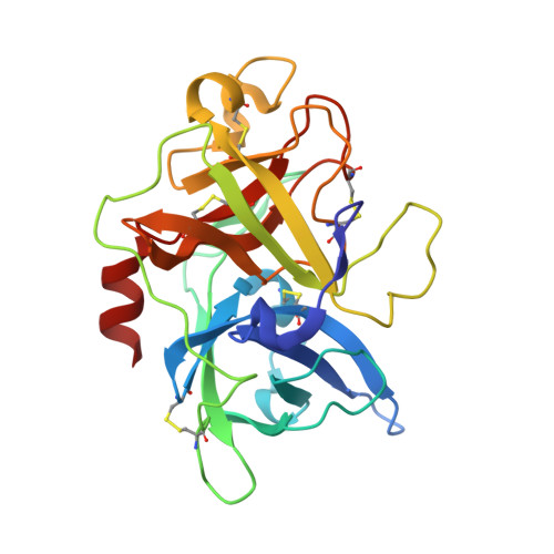
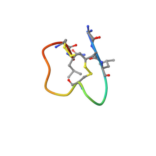
Crystal structure of urokinase-type plasminogen activator (uPA) complexed with bicyclic peptide UK604 (bicyclic 2)


Experimental Data Snapshot

  • Method: X-RAY DIFFRACTION
  • Resolution: 1.75 Å
  • R-Value Free: 0.186 
  • R-Value Work: 0.170 
  • R-Value Observed: 0.171 

wwPDB Validation   3D Report Full Report


This is version 1.3 of the entry. See complete history


Literature

Dithiol amino acids can structurally shape and enhance the ligand-binding properties of polypeptides.

Chen, S.Gopalakrishnan, R.Schaer, T.Marger, F.Hovius, R.Bertrand, D.Pojer, F.Heinis, C.

(2014) Nat Chem 6: 1009-1016

  • DOI: https://doi.org/10.1038/nchem.2043
  • Primary Citation of Related Structures:  
    2MOA, 4OS1, 4OS2, 4OS4, 4OS5, 4OS6, 4OS7

  • PubMed Abstract: 

    The disulfide bonds that form between two cysteine residues are important in defining and rigidifying the structures of proteins and peptides. In polypeptides containing multiple cysteine residues, disulfide isomerization can lead to multiple products with different biological activities. Here, we describe the development of a dithiol amino acid (Dtaa) that can form two disulfide bridges at a single amino acid site. Application of Dtaas to a serine protease inhibitor and a nicotinic acetylcholine receptor inhibitor that contain disulfide constraints enhanced their inhibitory activities 40- and 7.6-fold, respectively. X-ray crystallographic and NMR structure analysis show that the peptide ligands containing Dtaas have retained their native tertiary structures. We furthermore show that replacement of two cysteines by Dtaas can avoid the formation of disulfide bond isomers. With these properties, Dtaas are likely to have broad application in the rational design or directed evolution of peptides and proteins with high activity and stability.


  • Organizational Affiliation

    Institute of Chemical Sciences and Engineering, Ecole Polytechnique Fédérale de Lausanne, CH-1015 Lausanne, Switzerland.


Macromolecules
Find similar proteins by:  (by identity cutoff)  |  3D Structure
Entity ID: 1
MoleculeChains Sequence LengthOrganismDetailsImage
Urokinase-type plasminogen activator245Homo sapiensMutation(s): 2 
Gene Names: PLAU
EC: 3.4.21.73
UniProt & NIH Common Fund Data Resources
Find proteins for P00749 (Homo sapiens)
Explore P00749 
Go to UniProtKB:  P00749
PHAROS:  P00749
GTEx:  ENSG00000122861 
Entity Groups  
Sequence Clusters30% Identity50% Identity70% Identity90% Identity95% Identity100% Identity
UniProt GroupP00749
Sequence Annotations
Expand
  • Reference Sequence

Find similar proteins by:  Sequence   |   3D Structure  

Entity ID: 2
MoleculeChains Sequence LengthOrganismDetailsImage
bicyclic peptide UK604 (bicyclic 2)14N/AMutation(s): 0 
Entity Groups  
Sequence Clusters30% Identity50% Identity70% Identity90% Identity95% Identity100% Identity
Sequence Annotations
Expand
  • Reference Sequence
Biologically Interesting Molecules (External Reference) 1 Unique
Experimental Data & Validation

Experimental Data

  • Method: X-RAY DIFFRACTION
  • Resolution: 1.75 Å
  • R-Value Free: 0.186 
  • R-Value Work: 0.170 
  • R-Value Observed: 0.171 
  • Space Group: P 21 21 21
Unit Cell:
Length ( Å )Angle ( ˚ )
a = 52.35α = 90
b = 53.705β = 90
c = 84.659γ = 90
Software Package:
Software NamePurpose
PHASESphasing
REFMACrefinement
XDSdata reduction
SCALAdata scaling

Structure Validation

View Full Validation Report


Entry History 

Deposition Data

Revision History  (Full details and data files)

  • Version 1.0: 2014-09-24
    Type: Initial release
  • Version 1.1: 2014-11-05
    Changes: Database references
  • Version 1.2: 2017-11-22
    Changes: Refinement description
  • Version 1.3: 2021-06-02
    Changes: Database references, Derived calculations, Source and taxonomy