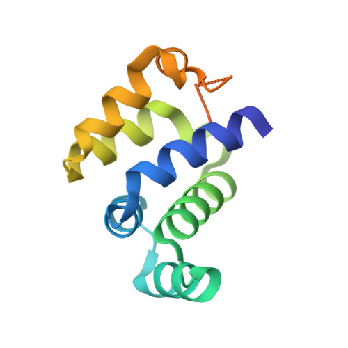
4O6X

Crystal structure of human Ankyrin G death domain


Experimental Data Snapshot

  • Method: X-RAY DIFFRACTION
  • Resolution: 2.10 Å
  • R-Value Free: 0.231 
  • R-Value Work: 0.182 
  • R-Value Observed: 0.185 

Starting Model: experimental
View more details

wwPDB Validation   3D Report Full Report


This is version 1.3 of the entry. See complete history


Literature

Crystal structure of human Ankyrin G death domain.

Liu, Y.Zhang, Y.Wang, J.H.

(2014) Proteins 82: 3476-3482

  • DOI: https://doi.org/10.1002/prot.24702
  • Primary Citation of Related Structures:  
    4O6X

  • PubMed Abstract: 

    Ankyrins (Ank) are a ubiquitously expressed family of multifunctional membrane adapter proteins. Ankyrin G (AnkG) is critical for assembling and maintenance of the axon initial segment. Here we present the 2.1 Å crystal structure of human AnkG death domain (hAnkG-DD). The core death domain is composed of six α-helices and three 3₁₀-helices. It forms a hydrophobic pocket on the surface of the molecule. The C-terminal tail of the hAnkG-DD curves back to have the aromatic ring of a phenylalanine residue, Phe100 insert into this pocket, which anchors the flexible tail onto the core domain. Related DDs were selected for structure comparison. The major variations are at the C-terminal region, including the α6 and the long C-terminal extension. The results of size exclusion chromatography and analytical ultracentrifugation suggest that hAnkG-DD exists as monomer in solution. Our work should help for the future investigation of the structure-function of AnkG.


  • Organizational Affiliation

    State Key Laboratory of Biomembrane and Membrane Biotechnology, College of Life Sciences, Peking University, Beijing, 100871, China.


Macromolecules
Find similar proteins by:  (by identity cutoff)  |  3D Structure
Entity ID: 1
MoleculeChains Sequence LengthOrganismDetailsImage
Ankyrin-3
A, B
121Homo sapiensMutation(s): 0 
Gene Names: ANK3Ankyrin G
UniProt & NIH Common Fund Data Resources
Find proteins for Q12955 (Homo sapiens)
Explore Q12955 
Go to UniProtKB:  Q12955
PHAROS:  Q12955
GTEx:  ENSG00000151150 
Entity Groups  
Sequence Clusters30% Identity50% Identity70% Identity90% Identity95% Identity100% Identity
UniProt GroupQ12955
Sequence Annotations
Expand
  • Reference Sequence
Experimental Data & Validation

Experimental Data

  • Method: X-RAY DIFFRACTION
  • Resolution: 2.10 Å
  • R-Value Free: 0.231 
  • R-Value Work: 0.182 
  • R-Value Observed: 0.185 
  • Space Group: P 21 21 21
Unit Cell:
Length ( Å )Angle ( ˚ )
a = 44.452α = 90
b = 56.817β = 90
c = 81.949γ = 90
Software Package:
Software NamePurpose
DENZOdata reduction
SCALEPACKdata scaling
MOLREPphasing
PHENIXrefinement
PDB_EXTRACTdata extraction
ADSCdata collection

Structure Validation

View Full Validation Report


Entry History 

Deposition Data

Revision History  (Full details and data files)

  • Version 1.0: 2014-10-29
    Type: Initial release
  • Version 1.1: 2014-12-17
    Changes: Database references
  • Version 1.2: 2015-02-11
    Changes: Other
  • Version 1.3: 2023-09-20
    Changes: Data collection, Database references, Refinement description