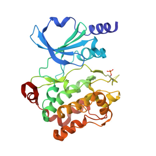
4O0X

Back pocket flexibility provides group-II PAK selectivity for type 1 kinase inhibitors


Experimental Data Snapshot

  • Method: X-RAY DIFFRACTION
  • Resolution: 2.48 Å
  • R-Value Free: 0.242 
  • R-Value Work: 0.203 
  • R-Value Observed: 0.205 

wwPDB Validation   3D Report Full Report


Ligand Structure Quality Assessment 


This is version 1.3 of the entry. See complete history


Literature

Back Pocket Flexibility Provides Group II p21-Activated Kinase (PAK) Selectivity for Type I 1/2 Kinase Inhibitors.

Staben, S.T.Feng, J.A.Lyle, K.Belvin, M.Boggs, J.Burch, J.D.Chua, C.C.Cui, H.Dipasquale, A.G.Friedman, L.S.Heise, C.Koeppen, H.Kotey, A.Mintzer, R.Oh, A.Roberts, D.A.Rouge, L.Rudolph, J.Tam, C.Wang, W.Xiao, Y.Young, A.Zhang, Y.Hoeflich, K.P.

(2014) J Med Chem 57: 1033-1045

  • DOI: https://doi.org/10.1021/jm401768t
  • Primary Citation of Related Structures:  
    4O0R, 4O0T, 4O0V, 4O0X, 4O0Y

  • PubMed Abstract: 

    Structure-based methods were used to design a potent and highly selective group II p21-activated kinase (PAK) inhibitor with a novel binding mode, compound 17. Hydrophobic interactions within a lipophilic pocket past the methionine gatekeeper of group II PAKs approached by these type I 1/2 binders were found to be important for improving potency. A structure-based hypothesis and strategy for achieving selectivity over group I PAKs, and the broad kinome, based on unique flexibility of this lipophilic pocket, is presented. A concentration-dependent decrease in tumor cell migration and invasion in two triple-negative breast cancer cell lines was observed with compound 17.


  • Organizational Affiliation

    Department of Discovery Chemistry, ‡Department of Translational Oncology, △Department of Pathology, §Department of Drug Metabolism and Pharmacokinetics, ∥Department of Biochemical and Cellular Pharmacology, and ⊥Department of Structural Biology, Genentech, Inc. , 1 DNA Way, South San Francisco, California 94080, United States.


Macromolecules
Find similar proteins by:  (by identity cutoff)  |  3D Structure
Entity ID: 1
MoleculeChains Sequence LengthOrganismDetailsImage
Serine/threonine-protein kinase PAK 4293Homo sapiensMutation(s): 0 
Gene Names: PAK4KIAA1142
EC: 2.7.11.1
UniProt & NIH Common Fund Data Resources
Find proteins for O96013 (Homo sapiens)
Explore O96013 
Go to UniProtKB:  O96013
PHAROS:  O96013
GTEx:  ENSG00000130669 
Entity Groups  
Sequence Clusters30% Identity50% Identity70% Identity90% Identity95% Identity100% Identity
UniProt GroupO96013
Sequence Annotations
Expand
  • Reference Sequence
Small Molecules
Ligands 1 Unique
IDChains Name / Formula / InChI Key2D Diagram3D Interactions
2OQ
Query on 2OQ

Download Ideal Coordinates CCD File 
B [auth A]1-{[1-(4-amino-1,3,5-triazin-2-yl)-2-methyl-1H-benzimidazol-6-yl]ethynyl}cyclohexanol
C19 H20 N6 O
RSIUBEYNBJJYTG-UHFFFAOYSA-N
Modified Residues  1 Unique
IDChains TypeFormula2D DiagramParent
SEP
Query on SEP
A
L-PEPTIDE LINKINGC3 H8 N O6 PSER
Binding Affinity Annotations 
IDSourceBinding Affinity
2OQ BindingDB:  4O0X Ki: 68 (nM) from 1 assay(s)
Experimental Data & Validation

Experimental Data

  • Method: X-RAY DIFFRACTION
  • Resolution: 2.48 Å
  • R-Value Free: 0.242 
  • R-Value Work: 0.203 
  • R-Value Observed: 0.205 
  • Space Group: P 41 21 2
Unit Cell:
Length ( Å )Angle ( ˚ )
a = 64.877α = 90
b = 64.877β = 90
c = 184.01γ = 90
Software Package:
Software NamePurpose
BOSdata collection
PHASERphasing
PHENIXrefinement
XDSdata reduction
SCALAdata scaling

Structure Validation

View Full Validation Report


Ligand Structure Quality Assessment 


Entry History 

Deposition Data

Revision History  (Full details and data files)

  • Version 1.0: 2014-02-12
    Type: Initial release
  • Version 1.1: 2014-02-26
    Changes: Database references
  • Version 1.2: 2014-11-05
    Changes: Structure summary
  • Version 1.3: 2024-10-09
    Changes: Data collection, Database references, Derived calculations, Structure summary