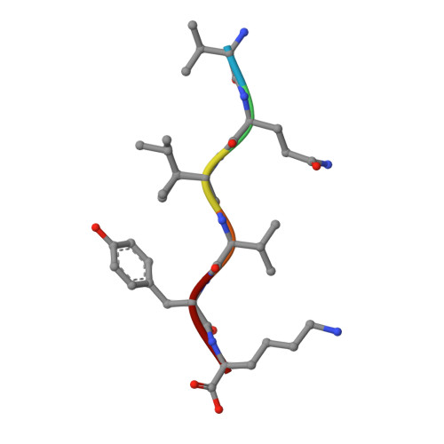
Molecular mechanisms for protein-encoded inheritance.
Wiltzius, J.J., Landau, M., Nelson, R., Sawaya, M.R., Apostol, M.I., Goldschmidt, L., Soriaga, A.B., Cascio, D., Rajashankar, K., Eisenberg, D.(2009) Nat Struct Mol Biol 16: 973-978
- PubMed: 19684598
- DOI: https://doi.org/10.1038/nsmb.1643
- Primary Citation of Related Structures:
3FOD, 3FPO, 3FR1, 3FTH, 3FTK, 3FTL, 3FTR, 3FVA, 4NP8 - PubMed Abstract:
In prion inheritance and transmission, strains are phenotypic variants encoded by protein 'conformations'. However, it is unclear how a protein conformation can be stable enough to endure transmission between cells or organisms. Here we describe new polymorphic crystal structures of segments of prion and other amyloid proteins, which offer two structural mechanisms for the encoding of prion strains. In packing polymorphism, prion strains are encoded by alternative packing arrangements (polymorphs) of beta-sheets formed by the same segment of a protein; in segmental polymorphism, prion strains are encoded by distinct beta-sheets built from different segments of a protein. Both forms of polymorphism can produce enduring conformations capable of encoding strains. These molecular mechanisms for transfer of protein-encoded information into prion strains share features with the familiar mechanism for transfer of nucleic acid-encoded information into microbial strains, including sequence specificity and recognition by noncovalent bonds.
Organizational Affiliation:
UCLA-DOE Institute for Genomics and Proteomics, Howard Hughes Medical Institute, Molecular Biology Institute, University of California, Los Angeles, California, USA.


