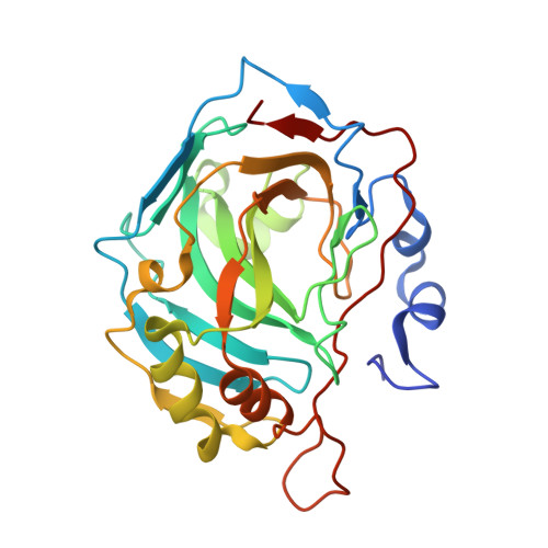
4L5W

Structural implications of the secondary CO2 binding pocket in human carbonic anhydrase II


Experimental Data Snapshot

  • Method: X-RAY DIFFRACTION
  • Resolution: 1.70 Å
  • R-Value Free: 0.181 
  • R-Value Work: 0.142 
  • R-Value Observed: 0.144 

wwPDB Validation   3D Report Full Report


This is version 1.1 of the entry. See complete history


Literature

Structural, catalytic and stabilizing consequences of aromatic cluster variants in human carbonic anhydrase II.

Boone, C.D.Gill, S.Tu, C.Silverman, D.N.McKenna, R.

(2013) Arch Biochem Biophys 539: 31-37

  • DOI: https://doi.org/10.1016/j.abb.2013.09.001
  • Primary Citation of Related Structures:  
    4L5U, 4L5V, 4L5W

  • PubMed Abstract: 

    The presence of aromatic clusters has been found to be an integral feature of many proteins isolated from thermophilic microorganisms. Residues found in aromatic cluster interact via π-π or C-H⋯π bonds between the phenyl rings, which are among the weakest interactions involved in protein stability. The lone aromatic cluster in human carbonic anhydrase II (HCA II) is centered on F226 with the surrounding aromatics F66, F95 and W97 located 12 Å posterior the active site; a location which could facilitate proper protein folding and active site construction. The role of F226 in the structure, catalytic activity and thermostability of HCA II was investigated via site-directed mutagenesis of three variants (F226I/L/W) into this position. The measured catalytic rates of the F226 variants via (18)O-mass spectrometry were identical to the native enzyme, but differential scanning calorimetry studies revealed a 3-4 K decrease in their denaturing temperature. X-ray crystallographic analysis suggests that the structural basis of this destabilization is via disruption and/or removal of weak C-H⋯π interactions between F226 to F66, F95 and W97. This study emphasizes the importance of the delicate arrangement of these weak interactions among aromatic clusters in overall protein stability.


  • Organizational Affiliation

    Biochemistry & Molecular Biology, University of Florida, P.O. Box 100245, Gainesville, FL 32610, United States.


Macromolecules
Find similar proteins by:  (by identity cutoff)  |  3D Structure
Entity ID: 1
MoleculeChains Sequence LengthOrganismDetailsImage
Carbonic anhydrase 2260Homo sapiensMutation(s): 2 
Gene Names: CA2
EC: 4.2.1.1 (PDB Primary Data), 4.2.1.69 (UniProt)
UniProt & NIH Common Fund Data Resources
Find proteins for P00918 (Homo sapiens)
Explore P00918 
Go to UniProtKB:  P00918
PHAROS:  P00918
GTEx:  ENSG00000104267 
Entity Groups  
Sequence Clusters30% Identity50% Identity70% Identity90% Identity95% Identity100% Identity
UniProt GroupP00918
Sequence Annotations
Expand
  • Reference Sequence
Small Molecules
Ligands 2 Unique
IDChains Name / Formula / InChI Key2D Diagram3D Interactions
GOL
Query on GOL

Download Ideal Coordinates CCD File 
C [auth A]GLYCEROL
C3 H8 O3
PEDCQBHIVMGVHV-UHFFFAOYSA-N
ZN
Query on ZN

Download Ideal Coordinates CCD File 
B [auth A]ZINC ION
Zn
PTFCDOFLOPIGGS-UHFFFAOYSA-N
Experimental Data & Validation

Experimental Data

  • Method: X-RAY DIFFRACTION
  • Resolution: 1.70 Å
  • R-Value Free: 0.181 
  • R-Value Work: 0.142 
  • R-Value Observed: 0.144 
  • Space Group: P 1 21 1
Unit Cell:
Length ( Å )Angle ( ˚ )
a = 42.117α = 90
b = 41.283β = 104.26
c = 72.02γ = 90
Software Package:
Software NamePurpose
PHENIXrefinement
PDB_EXTRACTdata extraction
CrystalCleardata collection
HKL-2000data reduction
HKL-2000data scaling
PHENIXphasing

Structure Validation

View Full Validation Report


Entry History 

Deposition Data

Revision History  (Full details and data files)

  • Version 1.0: 2014-04-30
    Type: Initial release
  • Version 1.1: 2024-02-28
    Changes: Data collection, Database references, Derived calculations