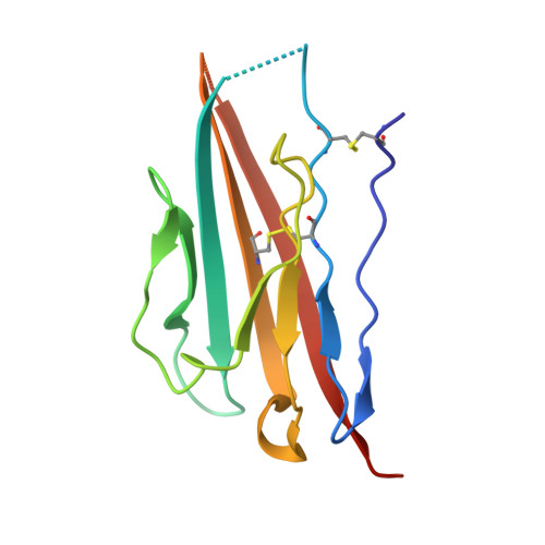
Crystal Structure and Molecular Imaging of the Nav Channel beta 3 Subunit Indicates a Trimeric Assembly.
Namadurai, S., Balasuriya, D., Rajappa, R., Wiemhofer, M., Stott, K., Klingauf, J., Edwardson, J.M., Chirgadze, D.Y., Jackson, A.P.(2014) J Biol Chem 289: 10797-10811
- PubMed: 24567321
- DOI: https://doi.org/10.1074/jbc.M113.527994
- Primary Citation of Related Structures:
4L1D - PubMed Abstract:
The vertebrate sodium (Nav) channel is composed of an ion-conducting α subunit and associated β subunits. Here, we report the crystal structure of the human β3 subunit immunoglobulin (Ig) domain, a functionally important component of Nav channels in neurons and cardiomyocytes. Surprisingly, we found that the β3 subunit Ig domain assembles as a trimer in the crystal asymmetric unit. Analytical ultracentrifugation confirmed the presence of Ig domain monomers, dimers, and trimers in free solution, and atomic force microscopy imaging also detected full-length β3 subunit monomers, dimers, and trimers. Mutation of a cysteine residue critical for maintaining the trimer interface destabilized both dimers and trimers. Using fluorescence photoactivated localization microscopy, we detected full-length β3 subunit trimers on the plasma membrane of transfected HEK293 cells. We further show that β3 subunits can bind to more than one site on the Nav 1.5 α subunit and induce the formation of α subunit oligomers, including trimers. Our results suggest a new and unexpected role for the β3 subunits in Nav channel cross-linking and provide new structural insights into some pathological Nav channel mutations.
Organizational Affiliation:
Department of Biochemistry, University of Cambridge, Tennis Court Road, Cambridge CB2 1QW, United Kingdom.