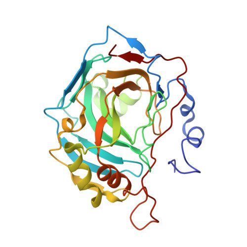
4KV0

Crystal structure of human carbonic anhydrase II in complex with the 5-(3-tosylureido)pyridine-2-sulfonamide inhibitor


Experimental Data Snapshot

  • Method: X-RAY DIFFRACTION
  • Resolution: 1.55 Å
  • R-Value Free: 0.179 
  • R-Value Work: 0.158 
  • R-Value Observed: 0.159 

Starting Model: experimental
View more details

wwPDB Validation   3D Report Full Report


Ligand Structure Quality Assessment 


This is version 1.2 of the entry. See complete history


Literature

Combining the tail and the ring approaches for obtaining potent and isoform-selective carbonic anhydrase inhibitors: solution and X-ray crystallographic studies.

Bozdag, M.Ferraroni, M.Nuti, E.Vullo, D.Rossello, A.Carta, F.Scozzafava, A.Supuran, C.T.

(2014) Bioorg Med Chem 22: 334-340

  • DOI: https://doi.org/10.1016/j.bmc.2013.11.016
  • Primary Citation of Related Structures:  
    4KV0

  • PubMed Abstract: 

    5-(3-Tosylureido)pyridine-2-sulfonamide and 4-tosylureido-benzenesulfonamide (ts-SA) only differ by the substitution of a CH by a nitrogen atom, but they have very different inhibitory properties against the metalloenzyme carbonic anhydrase (CA, EC 4.2.1.1). By means of X-ray crystallography on the human CA II adducts of the two compounds these differences have been rationalized. As all sulfonamides, the two compounds bind in deprotonated form to the Zn(II) ion from the enzyme active site and their organic scaffolds extend throughout the cavity, participating in many interactions with amino acid residues and water molecules. However the pyridine derivative undergoes a tilt of the heterocyclic ring compared to the benzene analog, which leads to a very different orientation of the two scaffolds when bound to the enzyme. This tilt also leads to a clash between a carbon atom from the pyridine ring of the first inhibitor and the OH moiety of Thr200, leading to less effective inhibitory properties of the pyridine versus the benzene sulfonamide derivative. Indeed, ts-SA is a promiscuous, low nanomolar inhibitor of 7 out of 10 human (h) CA isoforms, whereas the pyridine sulfonamide is a low nanomolar inhibitor only of the tumor-associated hCA IX and XII, being less effective against other 9 isoforms. Thus, a difference of one atom (N vs CH) in two isostructural sulfonamides leads to drastic differences of activity, phenomenon understood at the atomic level through the high resolution crystallographic structure and kinetic measurements reported in the paper. Combining the tail and the ring approaches in the same chemotype leads to isoform-selective, highly effective sulfonamide CA inhibitors.


  • Organizational Affiliation

    Università degli Studi di Firenze, Polo Scientifico, Laboratorio di Chimica Bioinorganica, Via della Lastruccia 3, 50019 Sesto Fiorentino (Florence), Italy.


Macromolecules
Find similar proteins by:  (by identity cutoff)  |  3D Structure
Entity ID: 1
MoleculeChains Sequence LengthOrganismDetailsImage
Carbonic anhydrase 2257Homo sapiensMutation(s): 0 
Gene Names: CA2
EC: 4.2.1.1 (PDB Primary Data), 4.2.1.69 (UniProt)
UniProt & NIH Common Fund Data Resources
Find proteins for P00918 (Homo sapiens)
Explore P00918 
Go to UniProtKB:  P00918
PHAROS:  P00918
GTEx:  ENSG00000104267 
Entity Groups  
Sequence Clusters30% Identity50% Identity70% Identity90% Identity95% Identity100% Identity
UniProt GroupP00918
Sequence Annotations
Expand
  • Reference Sequence
Small Molecules
Ligands 4 Unique
IDChains Name / Formula / InChI Key2D Diagram3D Interactions
MB7
Query on MB7

Download Ideal Coordinates CCD File 
E [auth A]5-({[(4-methylphenyl)sulfonyl]carbamoyl}amino)pyridine-2-sulfonamide
C13 H14 N4 O5 S2
PBRJWJGMKVRIRK-UHFFFAOYSA-N
GOL
Query on GOL

Download Ideal Coordinates CCD File 
C [auth A]GLYCEROL
C3 H8 O3
PEDCQBHIVMGVHV-UHFFFAOYSA-N
BME
Query on BME

Download Ideal Coordinates CCD File 
D [auth A]BETA-MERCAPTOETHANOL
C2 H6 O S
DGVVWUTYPXICAM-UHFFFAOYSA-N
ZN
Query on ZN

Download Ideal Coordinates CCD File 
B [auth A]ZINC ION
Zn
PTFCDOFLOPIGGS-UHFFFAOYSA-N
Experimental Data & Validation

Experimental Data

  • Method: X-RAY DIFFRACTION
  • Resolution: 1.55 Å
  • R-Value Free: 0.179 
  • R-Value Work: 0.158 
  • R-Value Observed: 0.159 
  • Space Group: P 1 21 1
Unit Cell:
Length ( Å )Angle ( ˚ )
a = 42.249α = 90
b = 41.283β = 104.28
c = 72.055γ = 90
Software Package:
Software NamePurpose
XSCALEdata scaling
REFMACrefinement
PDB_EXTRACTdata extraction
XDSdata reduction
XDSdata scaling

Structure Validation

View Full Validation Report


Ligand Structure Quality Assessment 


Entry History 

Deposition Data

Revision History  (Full details and data files)

  • Version 1.0: 2014-06-25
    Type: Initial release
  • Version 1.1: 2014-10-15
    Changes: Database references
  • Version 1.2: 2023-09-20
    Changes: Data collection, Database references, Derived calculations, Refinement description