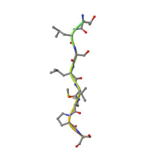
4KA3

Structure of MAP kinase in complex with a docking peptide


Experimental Data Snapshot

  • Method: X-RAY DIFFRACTION
  • Resolution: 2.71 Å
  • R-Value Free: 0.267 
  • R-Value Work: 0.225 
  • R-Value Observed: 0.227 

Starting Model: experimental
View more details

wwPDB Validation   3D Report Full Report


This is version 1.2 of the entry. See complete history


Literature

Crystal structure of the p38 alpha MAP kinase in complex with a docking peptide from TAB1

Xin, F.J.Wu, J.W.

(2013) Sci China Life Sci 56: 653-660

  • DOI: https://doi.org/10.1007/s11427-013-4494-0
  • Primary Citation of Related Structures:  
    4KA3

  • PubMed Abstract: 

    The mitogen-activated protein kinase (MAPK) p38α is a key regulator in many cellular processes, whose activity is tightly regulated by upstream kinases, phosphatases and other regulators. Transforming growth factor-β activated kinase 1 (TAK1) is an upstream kinase in p38α signaling, and its full activation requires a specific activator, the TAK1-binding protein (TAB1). TAB1 was also shown to be an inducer of p38α's autophosphorylation and/or a substrate driving the feedback control of p38α signaling. Here we determined the complex structure of the unphosphorylated p38α and a docking peptide of TAB1, which shows that the TAB1 peptide binds to the classical MAPK docking groove and induces long-range conformational changes on p38α. Our structural and biochemical analyses suggest that TAB1 is a reasonable substrate of p38α, yet the interaction between the docking peptide and p38α may not be sufficient to trigger trans-autophosphorylation of p38α.


  • Organizational Affiliation

    Tsinghua-Peking Center for Life Sciences and MOE Key Laboratory of Protein Science, School of Life Sciences, Tsinghua University, Beijing 100084, China.


Macromolecules
Find similar proteins by:  (by identity cutoff)  |  3D Structure
Entity ID: 1
MoleculeChains Sequence LengthOrganismDetailsImage
Mitogen-activated protein kinase 14360Mus musculusMutation(s): 0 
Gene Names: Mapk14
EC: 2.7.11.24
UniProt
Find proteins for P47811 (Mus musculus)
Explore P47811 
Go to UniProtKB:  P47811
Entity Groups  
Sequence Clusters30% Identity50% Identity70% Identity90% Identity95% Identity100% Identity
UniProt GroupP47811
Sequence Annotations
Expand
  • Reference Sequence
Find similar proteins by:  (by identity cutoff)  |  3D Structure
Entity ID: 2
MoleculeChains Sequence LengthOrganismDetailsImage
TGF-beta-activated kinase 1 and MAP3K7-binding protein 129Homo sapiensMutation(s): 0 
Gene Names: TAB1
UniProt & NIH Common Fund Data Resources
Find proteins for Q15750 (Homo sapiens)
Explore Q15750 
Go to UniProtKB:  Q15750
GTEx:  ENSG00000100324 
Entity Groups  
Sequence Clusters30% Identity50% Identity70% Identity90% Identity95% Identity100% Identity
UniProt GroupQ15750
Sequence Annotations
Expand
  • Reference Sequence
Experimental Data & Validation

Experimental Data

  • Method: X-RAY DIFFRACTION
  • Resolution: 2.71 Å
  • R-Value Free: 0.267 
  • R-Value Work: 0.225 
  • R-Value Observed: 0.227 
  • Space Group: P 31 2 1
Unit Cell:
Length ( Å )Angle ( ˚ )
a = 82.148α = 90
b = 82.148β = 90
c = 122.556γ = 120
Software Package:
Software NamePurpose
HKL-2000data collection
PHASERphasing
PHENIXrefinement
HKL-2000data reduction
HKL-2000data scaling

Structure Validation

View Full Validation Report


Entry History 

Deposition Data

Revision History  (Full details and data files)

  • Version 1.0: 2013-06-26
    Type: Initial release
  • Version 1.1: 2013-12-18
    Changes: Database references
  • Version 1.2: 2023-11-08
    Changes: Data collection, Database references, Refinement description