4GXB

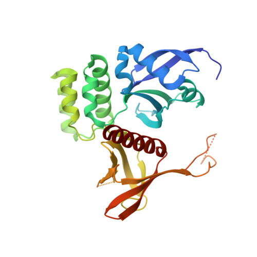
Structure of the SNX17 atypical FERM domain bound to the NPxY motif of P-selectin


Experimental Data Snapshot

  • Method: X-RAY DIFFRACTION
  • Resolution: 1.80 Å
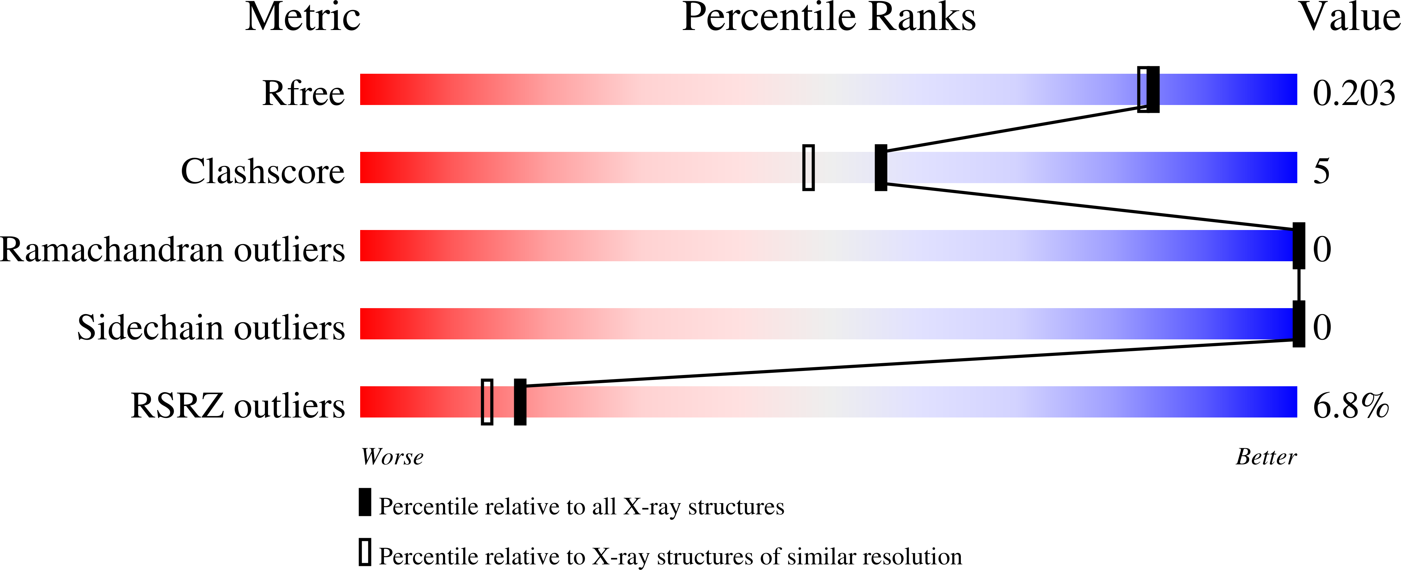
  • R-Value Free: 0.204 
  • R-Value Work: 0.177 
  • R-Value Observed: 0.178 

wwPDB Validation   3D Report Full Report


This is version 1.1 of the entry. See complete history


Literature

Structural basis for endosomal trafficking of diverse transmembrane cargos by PX-FERM proteins.

Ghai, R.Bugarcic, A.Liu, H.Norwood, S.J.Skeldal, S.Coulson, E.J.Li, S.S.Teasdale, R.D.Collins, B.M.

(2013) Proc Natl Acad Sci U S A 110: E643-E652

  • DOI: https://doi.org/10.1073/pnas.1216229110
  • Primary Citation of Related Structures:  
    4GXB

  • PubMed Abstract: 

    Transit of proteins through the endosomal organelle following endocytosis is critical for regulating the homeostasis of cell-surface proteins and controlling signal transduction pathways. However, the mechanisms that control these membrane-transport processes are poorly understood. The Phox-homology (PX) domain-containing proteins sorting nexin (SNX) 17, SNX27, and SNX31 have emerged recently as key regulators of endosomal recycling and bind conserved Asn-Pro-Xaa-Tyr-sorting signals in transmembrane cargos via an atypical band, 4.1/ezrin/radixin/moesin (FERM) domain. Here we present the crystal structure of the SNX17 FERM domain bound to the sorting motif of the P-selectin adhesion protein, revealing both the architecture of the atypical FERM domain and the molecular basis for recognition of these essential sorting sequences. We further show that the PX-FERM proteins share a promiscuous ability to bind a wide array of putative cargo molecules, including receptor tyrosine kinases, and propose a model for their coordinated molecular interactions with membrane, cargo, and regulatory proteins.


  • Organizational Affiliation

    Institute for Molecular Bioscience, The University of Queensland, St. Lucia, QLD 4072, Australia.


Macromolecules
Find similar proteins by:  (by identity cutoff)  |  3D Structure
Entity ID: 1
MoleculeChains Sequence LengthOrganismDetailsImage
Sorting nexin-17280Homo sapiensMutation(s): 0 
Gene Names: SNX17
UniProt & NIH Common Fund Data Resources
Find proteins for Q15036 (Homo sapiens)
Explore Q15036 
Go to UniProtKB:  Q15036
PHAROS:  Q15036
GTEx:  ENSG00000115234 
Entity Groups  
Sequence Clusters30% Identity50% Identity70% Identity90% Identity95% Identity100% Identity
UniProt GroupQ15036
Sequence Annotations
Expand
  • Reference Sequence
Find similar proteins by:  (by identity cutoff)  |  3D Structure
Entity ID: 2
MoleculeChains Sequence LengthOrganismDetailsImage
P-selectin41Mus musculusMutation(s): 0 
Gene Names: Selp
UniProt
Find proteins for Q01102 (Mus musculus)
Explore Q01102 
Go to UniProtKB:  Q01102
Entity Groups  
Sequence Clusters30% Identity50% Identity70% Identity90% Identity95% Identity100% Identity
UniProt GroupQ01102
Sequence Annotations
Expand
  • Reference Sequence
Small Molecules
Ligands 1 Unique
IDChains Name / Formula / InChI Key2D Diagram3D Interactions
GOL
Query on GOL

Download Ideal Coordinates CCD File 
C [auth A]GLYCEROL
C3 H8 O3
PEDCQBHIVMGVHV-UHFFFAOYSA-N
Experimental Data & Validation

Experimental Data

  • Method: X-RAY DIFFRACTION
  • Resolution: 1.80 Å
  • R-Value Free: 0.204 
  • R-Value Work: 0.177 
  • R-Value Observed: 0.178 
  • Space Group: P 31 2 1
Unit Cell:
Length ( Å )Angle ( ˚ )
a = 93.754α = 90
b = 93.754β = 90
c = 91.308γ = 120
Software Package:
Software NamePurpose
Blu-Icedata collection
SOLVEphasing
PHENIXrefinement
MOSFLMdata reduction
SCALAdata scaling

Structure Validation

View Full Validation Report


Entry History 

Deposition Data

Revision History  (Full details and data files)

  • Version 1.0: 2013-03-13
    Type: Initial release
  • Version 1.1: 2024-03-20
    Changes: Data collection, Database references, Derived calculations