4GEH

Crystal structure of MST4 dimerization domain complex with PDCD10


Experimental Data Snapshot

  • Method: X-RAY DIFFRACTION
  • Resolution: 1.95 Å
  • R-Value Free: 0.253 
  • R-Value Work: 0.220 
  • R-Value Observed: 0.222 

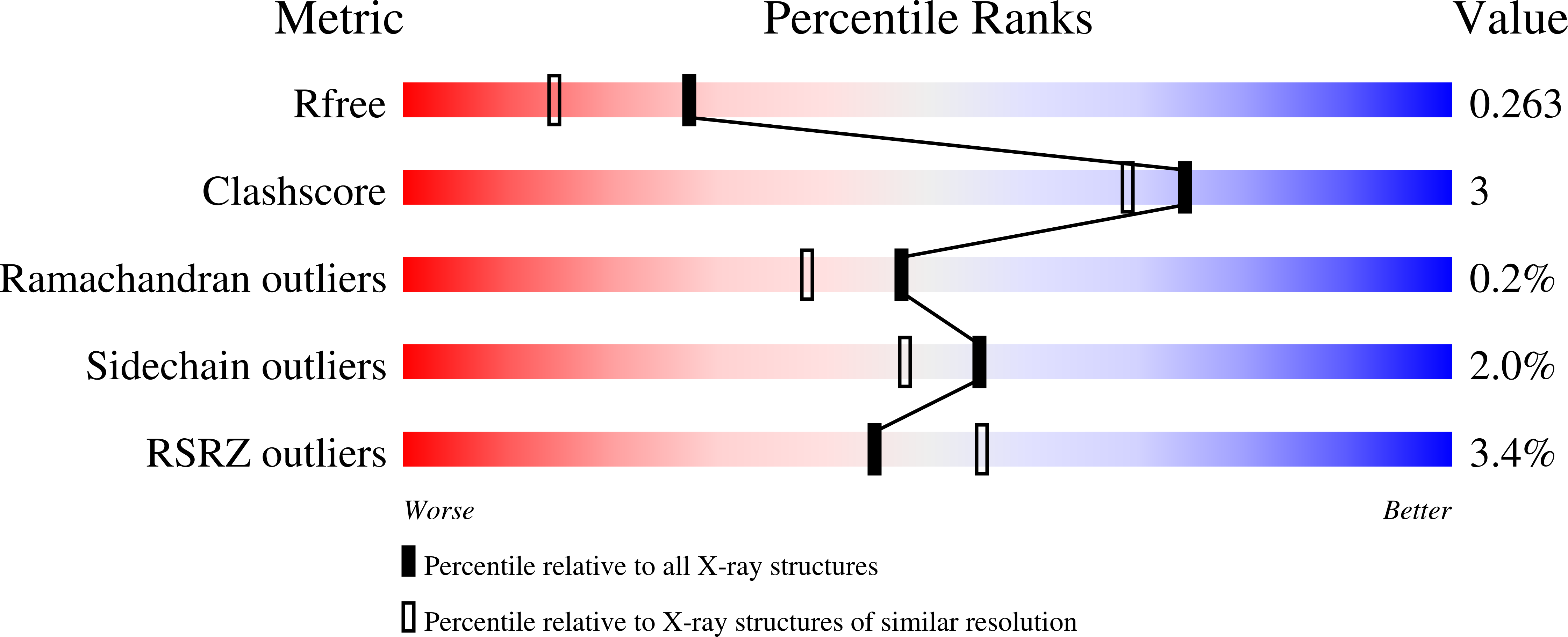
wwPDB Validation   3D Report Full Report


This is version 1.2 of the entry. See complete history


Literature

Structural mechanism of CCM3 heterodimerization with GCKIII kinases

Zhang, M.Dong, L.Shi, Z.Jiao, S.Zhang, Z.Zhang, W.Liu, G.Chen, C.Feng, M.Hao, Q.Wang, W.Yin, M.Zhao, Y.Zhang, L.Zhou, Z.

(2013) Structure 21: 680-688

  • DOI: https://doi.org/10.1016/j.str.2013.02.015
  • Primary Citation of Related Structures:  
    4GEH

  • PubMed Abstract: 

    Mutation of CCM3 causes cerebral cavernous malformations of the vasculature, leading to focal neurological deficits, seizures, and hemorrhagic stroke. CCM3 can heterodimerize with GCKIII kinases (MST3, MST4, and STK25) to regulate cardiovascular development. Here, we provide direct experimental evidence to prove that CCM3 heterodimerizes with GCKIII in a manner structurally resembling the CCM3 homodimerization. Structural comparison revealed the mechanism and critical residues that drive CCM3-GCKIII heterodimerization versus homodimerization. A flexible linker was identified for CCM3, which mediates a large-scale conformational rotation of the FAT domain relative to the dimerization domain. The conformational flip over of FAT domain removes steric locking in the CCM3 homodimer and allows its disassembly and subsequent heterodimerization with GCKIII. CCM3 forms a stable complex with MST4 in vivo to promote cell proliferation and migration synergistically in a manner dependent on MST4 kinase activity. Collectively, our work offers a structural basis for further functional study.


  • Organizational Affiliation

    State Key Laboratory of Cell Biology, Institute of Biochemistry and Cell Biology, Shanghai Institutes for Biological Sciences, Chinese Academy of Sciences, Shanghai 200031, China.


Macromolecules
Find similar proteins by:  (by identity cutoff)  |  3D Structure
Entity ID: 1
MoleculeChains Sequence LengthOrganismDetailsImage
Programmed cell death protein 10
A, C
207Homo sapiensMutation(s): 0 
Gene Names: PDCD10CCM3TFAR15
UniProt & NIH Common Fund Data Resources
Find proteins for Q9BUL8 (Homo sapiens)
Explore Q9BUL8 
Go to UniProtKB:  Q9BUL8
PHAROS:  Q9BUL8
GTEx:  ENSG00000114209 
Entity Groups  
Sequence Clusters30% Identity50% Identity70% Identity90% Identity95% Identity100% Identity
UniProt GroupQ9BUL8
Sequence Annotations
Expand
  • Reference Sequence
Find similar proteins by:  (by identity cutoff)  |  3D Structure
Entity ID: 2
MoleculeChains Sequence LengthOrganismDetailsImage
Serine/threonine-protein kinase MST4
B, D
91Homo sapiensMutation(s): 0 
Gene Names: MST4MASK
EC: 2.7.11.1
UniProt & NIH Common Fund Data Resources
Find proteins for Q9P289 (Homo sapiens)
Explore Q9P289 
Go to UniProtKB:  Q9P289
PHAROS:  Q9P289
GTEx:  ENSG00000134602 
Entity Groups  
Sequence Clusters30% Identity50% Identity70% Identity90% Identity95% Identity100% Identity
UniProt GroupQ9P289
Sequence Annotations
Expand
  • Reference Sequence
Experimental Data & Validation

Experimental Data

  • Method: X-RAY DIFFRACTION
  • Resolution: 1.95 Å
  • R-Value Free: 0.253 
  • R-Value Work: 0.220 
  • R-Value Observed: 0.222 
  • Space Group: P 21 21 21
Unit Cell:
Length ( Å )Angle ( ˚ )
a = 75.243α = 90
b = 84.018β = 90
c = 109.679γ = 90
Software Package:
Software NamePurpose
HKL-2000data collection
AutoSolphasing
REFMACrefinement
HKL-2000data reduction
HKL-2000data scaling

Structure Validation

View Full Validation Report


Entry History 

Deposition Data

Revision History  (Full details and data files)

  • Version 1.0: 2013-04-17
    Type: Initial release
  • Version 1.1: 2013-07-17
    Changes: Database references
  • Version 1.2: 2024-03-20
    Changes: Data collection, Database references, Refinement description