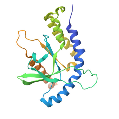
4F5W

Crystal structure of ligand free human STING CTD


Experimental Data Snapshot

  • Method: X-RAY DIFFRACTION
  • Resolution: 2.20 Å
  • R-Value Free: 0.275 
  • R-Value Work: 0.235 
  • R-Value Observed: 0.238 

wwPDB Validation   3D Report Full Report


This is version 1.2 of the entry. See complete history


Literature

Crystal structures of STING protein reveal basis for recognition of cyclic di-GMP

Shang, G.Zhu, D.Li, N.Zhang, J.Zhu, C.Lu, D.Liu, C.Yu, Q.Zhao, Y.Xu, S.Gu, L.

(2012) Nat Struct Mol Biol 19: 725-727

  • DOI: https://doi.org/10.1038/nsmb.2332
  • Primary Citation of Related Structures:  
    4F5W, 4F5Y

  • PubMed Abstract: 

    STING functions as both an adaptor protein signaling cytoplasmic double-stranded DNA and a direct immunosensor of cyclic diguanylate monophosphate (c-di-GMP). The crystal structures of the C-terminal domain of human STING (STING(CTD)) and its complex with c-di-GMP reveal how STING recognizes c-di-GMP. In response to c-di-GMP binding, two surface loops, which serve as a gate and latch of the cleft formed by the dimeric STING(CTD), undergo rearrangements to interact with the ligand.


  • Organizational Affiliation

    State Key Laboratory of Microbial Technology, School of Life Science, Shandong University, Jinan, China.


Macromolecules
Find similar proteins by:  (by identity cutoff)  |  3D Structure
Entity ID: 1
MoleculeChains Sequence LengthOrganismDetailsImage
Transmembrane protein 173239Homo sapiensMutation(s): 0 
Gene Names: TMEM173ERISMITASTING
UniProt & NIH Common Fund Data Resources
Find proteins for Q86WV6 (Homo sapiens)
Explore Q86WV6 
Go to UniProtKB:  Q86WV6
PHAROS:  Q86WV6
GTEx:  ENSG00000184584 
Entity Groups  
Sequence Clusters30% Identity50% Identity70% Identity90% Identity95% Identity100% Identity
UniProt GroupQ86WV6
Sequence Annotations
Expand
  • Reference Sequence
Small Molecules
Ligands 1 Unique
IDChains Name / Formula / InChI Key2D Diagram3D Interactions
CA
Query on CA

Download Ideal Coordinates CCD File 
B [auth A]CALCIUM ION
Ca
BHPQYMZQTOCNFJ-UHFFFAOYSA-N
Experimental Data & Validation

Experimental Data

  • Method: X-RAY DIFFRACTION
  • Resolution: 2.20 Å
  • R-Value Free: 0.275 
  • R-Value Work: 0.235 
  • R-Value Observed: 0.238 
  • Space Group: C 2 2 21
Unit Cell:
Length ( Å )Angle ( ˚ )
a = 81.099α = 90
b = 91.028β = 90
c = 73.395γ = 90
Software Package:
Software NamePurpose
HKL-2000data collection
SOLVEphasing
PHENIXrefinement
HKL-2000data reduction
HKL-2000data scaling

Structure Validation

View Full Validation Report


Entry History 

Deposition Data

Revision History  (Full details and data files)

  • Version 1.0: 2012-06-27
    Type: Initial release
  • Version 1.1: 2013-09-04
    Changes: Database references
  • Version 1.2: 2024-03-20
    Changes: Data collection, Database references, Derived calculations