4EKD

Structure of human regulator of G protein signaling 2 (RGS2) in complex with murine Galpha-q(R183C)


Experimental Data Snapshot

  • Method: X-RAY DIFFRACTION
  • Resolution: 2.71 Å
  • R-Value Free: 0.252 
  • R-Value Work: 0.190 
  • R-Value Observed: 0.193 

Starting Models: experimental
View more details

wwPDB Validation   3D Report Full Report


Ligand Structure Quality Assessment 


This is version 1.3 of the entry. See complete history


Literature

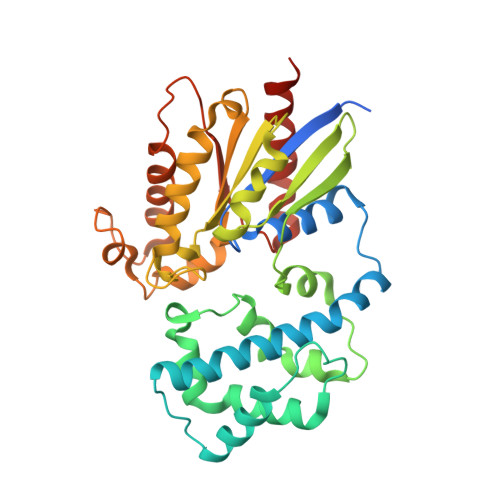
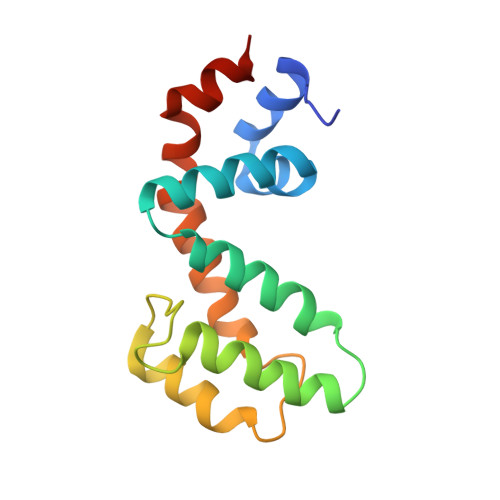
Structural and functional analysis of the regulator of G protein signaling 2-g alpha q complex.

Nance, M.R.Kreutz, B.Tesmer, V.M.Sterne-Marr, R.Kozasa, T.Tesmer, J.J.

(2013) Structure 21: 438-448

  • DOI: https://doi.org/10.1016/j.str.2012.12.016
  • Primary Citation of Related Structures:  
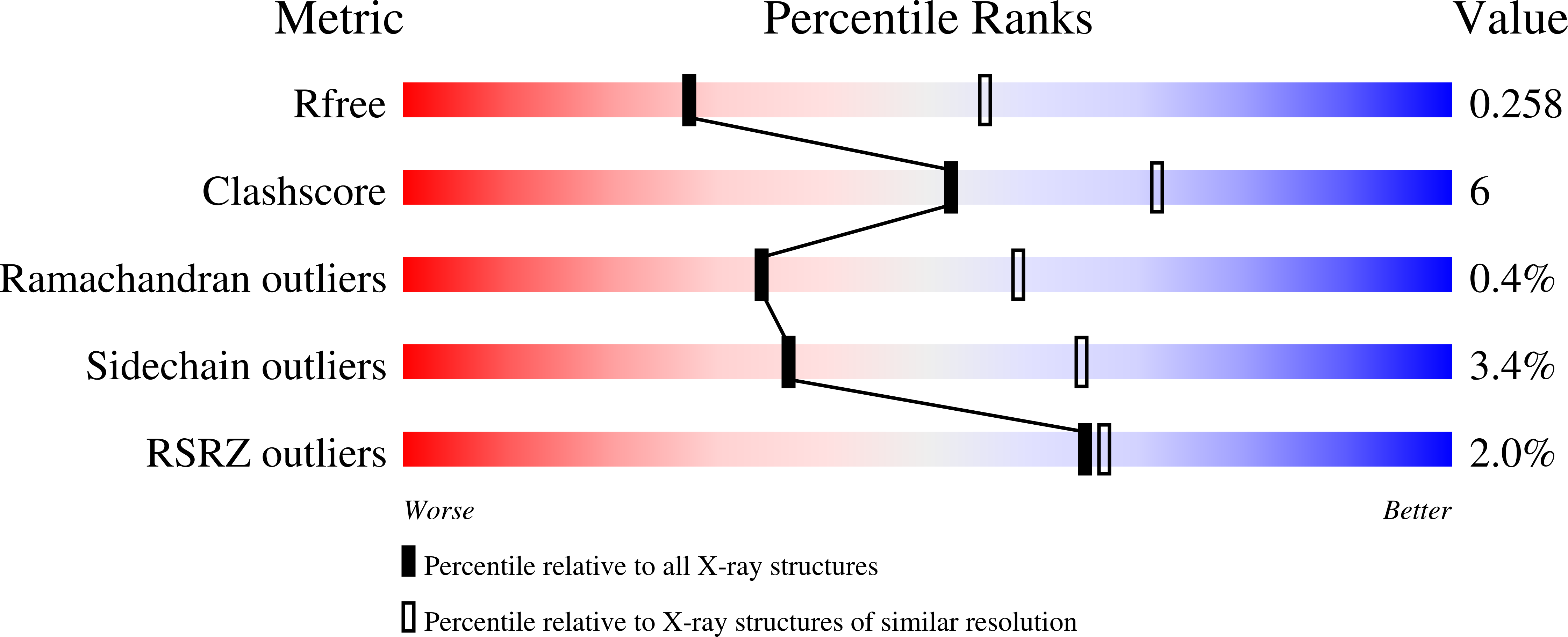
    4EKC, 4EKD

  • PubMed Abstract: 

    The heterotrimeric G protein Gαq is a key regulator of blood pressure, and excess Gαq signaling leads to hypertension. A specific inhibitor of Gαq is the GTPase activating protein (GAP) known as regulator of G protein signaling 2 (RGS2). The molecular basis for how Gαq/11 subunits serve as substrates for RGS proteins and how RGS2 mandates its selectivity for Gαq is poorly understood. In crystal structures of the RGS2-Gαq complex, RGS2 docks to Gαq in a different orientation from that observed in RGS-Gαi/o complexes. Despite its unique pose, RGS2 maintains canonical interactions with the switch regions of Gαq in part because its α6 helix adopts a distinct conformation. We show that RGS2 forms extensive interactions with the α-helical domain of Gαq that contribute to binding affinity and GAP potency. RGS subfamilies that do not serve as GAPs for Gαq are unlikely to form analogous stabilizing interactions.


  • Organizational Affiliation

    Life Sciences Institute, University of Michigan, Ann Arbor, MI 48109-2216, USA.


Macromolecules
Find similar proteins by:  (by identity cutoff)  |  3D Structure
Entity ID: 1
MoleculeChains Sequence LengthOrganismDetailsImage
Guanine nucleotide-binding protein G(q) subunit alpha347Mus musculusMutation(s): 6 
Gene Names: Gnaq
EC: 3.6.5.1
UniProt
Find proteins for P21279 (Mus musculus)
Explore P21279 
Go to UniProtKB:  P21279
Entity Groups  
Sequence Clusters30% Identity50% Identity70% Identity90% Identity95% Identity100% Identity
UniProt GroupP21279
Sequence Annotations
Expand
  • Reference Sequence
Find similar proteins by:  (by identity cutoff)  |  3D Structure
Entity ID: 2
MoleculeChains Sequence LengthOrganismDetailsImage
Regulator of G-protein signaling 2137Homo sapiensMutation(s): 0 
Gene Names: RGS2G0S8GIG31
UniProt & NIH Common Fund Data Resources
Find proteins for P41220 (Homo sapiens)
Explore P41220 
Go to UniProtKB:  P41220
GTEx:  ENSG00000116741 
Entity Groups  
Sequence Clusters30% Identity50% Identity70% Identity90% Identity95% Identity100% Identity
UniProt GroupP41220
Sequence Annotations
Expand
  • Reference Sequence
Small Molecules
Ligands 6 Unique
IDChains Name / Formula / InChI Key2D Diagram3D Interactions
GDP
Query on GDP

Download Ideal Coordinates CCD File 
C [auth A]GUANOSINE-5'-DIPHOSPHATE
C10 H15 N5 O11 P2
QGWNDRXFNXRZMB-UUOKFMHZSA-N
MES
Query on MES

Download Ideal Coordinates CCD File 
J [auth B]2-(N-MORPHOLINO)-ETHANESULFONIC ACID
C6 H13 N O4 S
SXGZJKUKBWWHRA-UHFFFAOYSA-N
ALF
Query on ALF

Download Ideal Coordinates CCD File 
D [auth A]TETRAFLUOROALUMINATE ION
Al F4
UYOMQIYKOOHAMK-UHFFFAOYSA-J
CO
Query on CO

Download Ideal Coordinates CCD File 
E [auth A],
H [auth A]
COBALT (II) ION
Co
XLJKHNWPARRRJB-UHFFFAOYSA-N
CL
Query on CL

Download Ideal Coordinates CCD File 
F [auth A],
G [auth A]
CHLORIDE ION
Cl
VEXZGXHMUGYJMC-UHFFFAOYSA-M
MG
Query on MG

Download Ideal Coordinates CCD File 
I [auth A]MAGNESIUM ION
Mg
JLVVSXFLKOJNIY-UHFFFAOYSA-N
Experimental Data & Validation

Experimental Data

  • Method: X-RAY DIFFRACTION
  • Resolution: 2.71 Å
  • R-Value Free: 0.252 
  • R-Value Work: 0.190 
  • R-Value Observed: 0.193 
  • Space Group: P 41 21 2
Unit Cell:
Length ( Å )Angle ( ˚ )
a = 60.248α = 90
b = 60.248β = 90
c = 345.569γ = 90
Software Package:
Software NamePurpose
PHASERphasing
REFMACrefinement
HKL-2000data reduction
HKL-2000data scaling

Structure Validation

View Full Validation Report


Ligand Structure Quality Assessment 


Entry History 

Deposition Data

Revision History  (Full details and data files)

  • Version 1.0: 2013-01-30
    Type: Initial release
  • Version 1.1: 2013-08-28
    Changes: Database references
  • Version 1.2: 2019-11-20
    Changes: Database references, Derived calculations
  • Version 1.3: 2023-09-13
    Changes: Data collection, Database references, Derived calculations, Refinement description