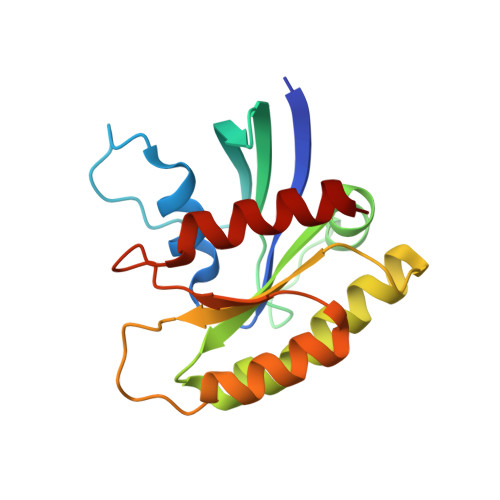
4EFM

Crystal structure of H-Ras G12V in complex with GppNHp (state 1)


Experimental Data Snapshot

  • Method: X-RAY DIFFRACTION
  • Resolution: 1.90 Å
  • R-Value Free: 0.221 
  • R-Value Work: 0.186 
  • R-Value Observed: 0.188 

Starting Model: experimental
View more details

wwPDB Validation   3D Report Full Report


Ligand Structure Quality Assessment 


This is version 1.2 of the entry. See complete history


Literature

Crystal structures of the state 1 conformations of the GTP-bound H-Ras protein and its oncogenic G12V and Q61L mutants

Muraoka, S.Shima, F.Araki, M.Inoue, T.Yoshimoto, A.Ijiri, Y.Seki, N.Tamura, A.Kumasaka, T.Yamamoto, M.Kataoka, T.

(2012) FEBS Lett 586: 1715-1718

  • DOI: https://doi.org/10.1016/j.febslet.2012.04.058
  • Primary Citation of Related Structures:  
    4EFL, 4EFM, 4EFN

  • PubMed Abstract: 

    GTP-bound Ras adopts two interconverting conformations, "inactive" state 1 and "active" state 2. However, the tertiary structure of wild-type (WT) state 1 remains unsolved. Here we solve the state 1 crystal structures of H-Ras WT together with its oncogenic G12V and Q61L mutants. They assume open structures characterized by impaired interactions of both Thr-35 in switch I and Gly-60 in switch II with the γ-phosphate of GTP and possess two surface pockets of mutually different shapes unseen in state 2, a potential target for selective inhibitor development. Furthermore, they provide a structural basis for the low GTPase activity of state 1.


  • Organizational Affiliation

    Division of Molecular Biology, Department of Biochemistry and Molecular Biology, Kobe University Graduate School of Medicine, 7-5-1 Kusunoki-cho, Chuo-ku, Kobe 650-0017, Japan.


Macromolecules
Find similar proteins by:  (by identity cutoff)  |  3D Structure
Entity ID: 1
MoleculeChains Sequence LengthOrganismDetailsImage
GTPase HRas171Homo sapiensMutation(s): 1 
Gene Names: HRASHRAS1
EC: 3.6.5.2
UniProt & NIH Common Fund Data Resources
Find proteins for P01112 (Homo sapiens)
Explore P01112 
Go to UniProtKB:  P01112
PHAROS:  P01112
GTEx:  ENSG00000174775 
Entity Groups  
Sequence Clusters30% Identity50% Identity70% Identity90% Identity95% Identity100% Identity
UniProt GroupP01112
Sequence Annotations
Expand
  • Reference Sequence
Small Molecules
Ligands 2 Unique
IDChains Name / Formula / InChI Key2D Diagram3D Interactions
GNP
Query on GNP

Download Ideal Coordinates CCD File 
B [auth A]PHOSPHOAMINOPHOSPHONIC ACID-GUANYLATE ESTER
C10 H17 N6 O13 P3
UQABYHGXWYXDTK-UUOKFMHZSA-N
MG
Query on MG

Download Ideal Coordinates CCD File 
C [auth A]MAGNESIUM ION
Mg
JLVVSXFLKOJNIY-UHFFFAOYSA-N
Experimental Data & Validation

Experimental Data

  • Method: X-RAY DIFFRACTION
  • Resolution: 1.90 Å
  • R-Value Free: 0.221 
  • R-Value Work: 0.186 
  • R-Value Observed: 0.188 
  • Space Group: I 2 2 2
Unit Cell:
Length ( Å )Angle ( ˚ )
a = 34.638α = 90
b = 81.833β = 90
c = 121.761γ = 90
Software Package:
Software NamePurpose
BSSdata collection
REFMACrefinement
MOSFLMdata reduction
SCALAdata scaling

Structure Validation

View Full Validation Report


Ligand Structure Quality Assessment 


Entry History 

Deposition Data

Revision History  (Full details and data files)

  • Version 1.0: 2012-05-16
    Type: Initial release
  • Version 1.1: 2014-03-12
    Changes: Database references
  • Version 1.2: 2023-11-08
    Changes: Data collection, Database references, Derived calculations, Refinement description