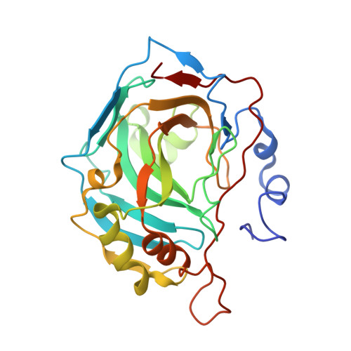
4BF1

Three dimensional structure of human carbonic anhydrase II in complex with 5-(1-naphthalen-1-yl-1,2,3-triazol-4-yl)thiophene-2-sulfonamide


Experimental Data Snapshot

  • Method: X-RAY DIFFRACTION
  • Resolution: 1.35 Å
  • R-Value Free: 0.181 
  • R-Value Work: 0.127 
  • R-Value Observed: 0.130 

Starting Model: experimental
View more details

wwPDB Validation   3D Report Full Report


Ligand Structure Quality Assessment 


This is version 1.4 of the entry. See complete history


Literature

5-Substituted-(1,2,3-Triazol-4-Yl)Thiophene-2-Sulfonamides Strongly Inhibit Human Carbonic Anhydrases I, II, Ix and Xii: Solution and X-Ray Crystallographic Studies.

Leitans, J.Sprudza, A.Tanc, M.Vozny, I.Zalubovskis, R.Tars, K.Supuran, C.T.

(2013) Bioorg Med Chem 21: 5130

  • DOI: https://doi.org/10.1016/j.bmc.2013.06.041
  • Primary Citation of Related Structures:  
    4BF1, 4BF6

  • PubMed Abstract: 

    We report here a series of 2-thiophene-sulfonamides incorporating 1-substituted aryl-1,2,3-triazolyl moieties, prepared by click chemistry from 5-ethynylthiophene-2-sulfonamide and substituted aryl azides. The new sulfonamides were investigated as inhibitors of the zinc metalloenzyme CA (EC 4.2.1.1), and more specifically against the human (h) cytosolic isoforms hCA I and II and the transmembrane, tumor-associated ones hCA IX and XII: The new compounds were medium-weak hCA I inhibitors (KIs in the range of 224-7544nM), but were compactly, highly effective, low nanomolar hCA II inhibitors (KIs of 2.2-7.7nM). The tumor-associated hCA IX was inhibited with KIs ranging between 5.4 and 811nM, whereas hCA XII with inhibition constants in the range of 3.4-239nM. The X-ray crystal structure of the adducts of two such compounds bound to hCA II (one incorporating 1-naphthyl, the other one 3-cyanophenyl moieties) evidenced the reasons of the high affinity for hCA II. Highly favorable, predominantly hydrophobic interactions between the sulfonamide scaffold and the hCA II active site were responsible for the binding, in addition to the coordination of the sulfamoyl moiety to the zinc ion. The tails of the two inhibitors adopted very diverse orientations when bound to the active site, with the naphthyltriazolyl moiety orientated towards the hydrophobic half of the active site, and the 3-cyanophenyl one pointing towards the hydrophilic half. These data may be used for the structure-based drug design of even more effective hCA II inhibitors, with potential use as antiglaucoma agents or as diuretics.


  • Organizational Affiliation

    Biomedical Research and Study Center, Ratsupites 1, LV 1067 Riga, Latvia.


Macromolecules
Find similar proteins by:  (by identity cutoff)  |  3D Structure
Entity ID: 1
MoleculeChains Sequence LengthOrganismDetailsImage
CARBONIC ANHYDRASE 2259Homo sapiensMutation(s): 0 
EC: 4.2.1.1 (PDB Primary Data), 4.2.1.69 (UniProt)
UniProt & NIH Common Fund Data Resources
Find proteins for P00918 (Homo sapiens)
Explore P00918 
Go to UniProtKB:  P00918
PHAROS:  P00918
GTEx:  ENSG00000104267 
Entity Groups  
Sequence Clusters30% Identity50% Identity70% Identity90% Identity95% Identity100% Identity
UniProt GroupP00918
Sequence Annotations
Expand
  • Reference Sequence
Small Molecules
Ligands 3 Unique
IDChains Name / Formula / InChI Key2D Diagram3D Interactions
9FK
Query on 9FK

Download Ideal Coordinates CCD File 
C [auth A]5-(1-naphthalen-1-yl-1,2,3-triazol-4-yl)thiophene-2-sulfonamide
C16 H12 N4 O2 S2
KNFVNHRBTIJTNM-UHFFFAOYSA-N
ZN
Query on ZN

Download Ideal Coordinates CCD File 
B [auth A]ZINC ION
Zn
PTFCDOFLOPIGGS-UHFFFAOYSA-N
NA
Query on NA

Download Ideal Coordinates CCD File 
D [auth A]SODIUM ION
Na
FKNQFGJONOIPTF-UHFFFAOYSA-N
Experimental Data & Validation

Experimental Data

  • Method: X-RAY DIFFRACTION
  • Resolution: 1.35 Å
  • R-Value Free: 0.181 
  • R-Value Work: 0.127 
  • R-Value Observed: 0.130 
  • Space Group: P 1 21 1
Unit Cell:
Length ( Å )Angle ( ˚ )
a = 42.32α = 90
b = 41.53β = 104.28
c = 72.21γ = 90
Software Package:
Software NamePurpose
REFMACrefinement
MOSFLMdata reduction
SCALAdata scaling

Structure Validation

View Full Validation Report


Ligand Structure Quality Assessment 


Entry History 

Deposition Data

Revision History  (Full details and data files)

  • Version 1.0: 2014-01-22
    Type: Initial release
  • Version 1.1: 2018-01-17
    Changes: Data collection
  • Version 1.2: 2019-01-30
    Changes: Data collection, Experimental preparation
  • Version 1.3: 2019-02-06
    Changes: Data collection, Experimental preparation
  • Version 1.4: 2023-12-20
    Changes: Data collection, Database references, Derived calculations, Other, Refinement description