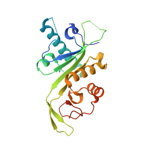
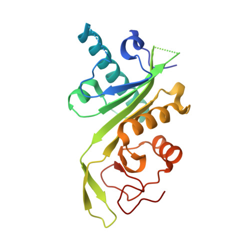
4B5P

Crystal structure of human alpha tubulin acetyltransferase catalytic domain Q58A variant


Experimental Data Snapshot

  • Method: X-RAY DIFFRACTION
  • Resolution: 1.60 Å
  • R-Value Free: 0.222 
  • R-Value Work: 0.185 
  • R-Value Observed: 0.186 

Starting Model: experimental
View more details

wwPDB Validation   3D Report Full Report


Ligand Structure Quality Assessment 


This is version 1.3 of the entry. See complete history


Literature

Atomic Resolution Structure of Human Alpha-Tubulin Acetyltransferase Bound to Acetyl-Coa.

Taschner, M.Vetter, M.Lorentzen, E.

(2012) Proc Natl Acad Sci U S A 109: 19649

  • DOI: https://doi.org/10.1073/pnas.1209343109
  • Primary Citation of Related Structures:  
    4B5O, 4B5P

  • PubMed Abstract: 

    Acetylation of lysine residues is an important posttranslational modification found in all domains of life. α-Tubulin is specifically acetylated on lysine 40, a modification that serves to stabilize microtubules of axons and cilia. Whereas histone acetyltransferases have been extensively studied, there is no structural and mechanistic information available on α-tubulin acetyltransferases. Here, we present the structure of the human α-tubulin acetyltransferase catalytic domain bound to its cosubstrate acetyl-CoA at 1.05 Å resolution. Compared with other lysine acetyltransferases of known structure, α-tubulin acetyltransferase displays a relatively well-conserved cosubstrate binding pocket but is unique in its active site and putative α-tubulin binding site. Using acetylation assays with structure-guided mutants, we map residues important for acetyl-CoA binding, substrate binding, and catalysis. This analysis reveals a basic patch implicated in substrate binding and a conserved glutamine residue required for catalysis, demonstrating that the family of α-tubulin acetyltransferases uses a reaction mechanism different from other lysine acetyltransferases characterized to date.


  • Organizational Affiliation

    Department of Structural Cell Biology, Max Planck Institute of Biochemistry, D-82152 Martinsried, Germany.


Macromolecules
Find similar proteins by:  (by identity cutoff)  |  3D Structure
Entity ID: 1
MoleculeChains Sequence LengthOrganismDetailsImage
ALPHA-TUBULIN N-ACETYLTRANSFERASE200Homo sapiensMutation(s): 1 
EC: 2.3.1.108
UniProt & NIH Common Fund Data Resources
Find proteins for Q5SQI0 (Homo sapiens)
Explore Q5SQI0 
Go to UniProtKB:  Q5SQI0
GTEx:  ENSG00000137343 
Entity Groups  
Sequence Clusters30% Identity50% Identity70% Identity90% Identity95% Identity100% Identity
UniProt GroupQ5SQI0
Sequence Annotations
Expand
  • Reference Sequence
Find similar proteins by:  (by identity cutoff)  |  3D Structure
Entity ID: 2
MoleculeChains Sequence LengthOrganismDetailsImage
ALPHA-TUBULIN N-ACETYLTRANSFERASE200Homo sapiensMutation(s): 1 
EC: 2.3.1.108
UniProt & NIH Common Fund Data Resources
Find proteins for Q5SQI0 (Homo sapiens)
Explore Q5SQI0 
Go to UniProtKB:  Q5SQI0
GTEx:  ENSG00000137343 
Entity Groups  
Sequence Clusters30% Identity50% Identity70% Identity90% Identity95% Identity100% Identity
UniProt GroupQ5SQI0
Sequence Annotations
Expand
  • Reference Sequence
Experimental Data & Validation

Experimental Data

  • Method: X-RAY DIFFRACTION
  • Resolution: 1.60 Å
  • R-Value Free: 0.222 
  • R-Value Work: 0.185 
  • R-Value Observed: 0.186 
  • Space Group: P 21 21 21
Unit Cell:
Length ( Å )Angle ( ˚ )
a = 36.76α = 90
b = 110.87β = 90
c = 113.58γ = 90
Software Package:
Software NamePurpose
PHENIXrefinement
XDSdata reduction
XDSdata scaling
PHASERphasing

Structure Validation

View Full Validation Report


Ligand Structure Quality Assessment 


Entry History 

Deposition Data

Revision History  (Full details and data files)

  • Version 1.0: 2012-10-24
    Type: Initial release
  • Version 1.1: 2012-12-12
    Changes: Database references
  • Version 1.2: 2013-09-11
    Changes: Atomic model, Database references, Other, Source and taxonomy, Structure summary
  • Version 1.3: 2023-12-20
    Changes: Data collection, Database references, Derived calculations, Other, Refinement description