3W51

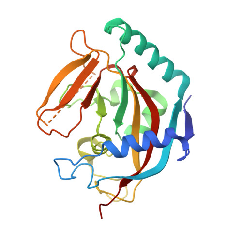
Tankyrase in complex with 2-hydroxy-4-methylquinoline


Experimental Data Snapshot

  • Method: X-RAY DIFFRACTION
  • Resolution: 2.00 Å
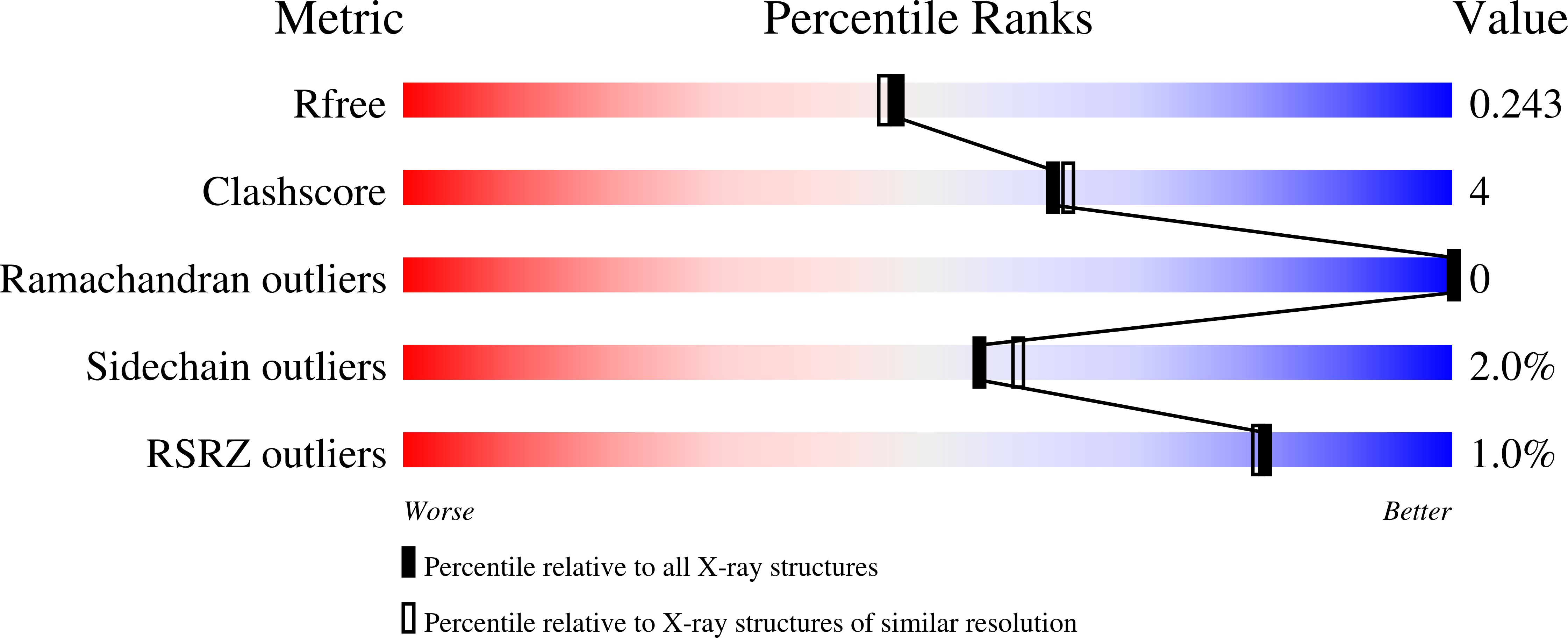
  • R-Value Free: 0.243 
  • R-Value Work: 0.187 
  • R-Value Observed: 0.190 

Starting Model: experimental
View more details

wwPDB Validation   3D Report Full Report


Ligand Structure Quality Assessment 


This is version 1.1 of the entry. See complete history


Literature

Fragment-based ligand design of novel potent inhibitors of tankyrases.

Larsson, E.A.Jansson, A.E.Ng, F.M.Then, S.W.Panicker, R.Liu, B.Sangthongpitag, K.Pendharkar, V.Tai, S.J.Hill, J.Dan, C.Ho, S.Y.Cheong, W.W.Poulsen, A.Blanchard, S.Lin, G.R.Alam, J.Keller, T.H.Nordlund, P.

(2013) J Med Chem 56: 4497-4508

  • DOI: https://doi.org/10.1021/jm400211f
  • Primary Citation of Related Structures:  
    3W51, 4IUE, 4J1Z, 4J21, 4J22, 4J3L, 4J3M

  • PubMed Abstract: 

    Tankyrases constitute potential drug targets for cancer and myelin-degrading diseases. We have applied a structure- and biophysics-driven fragment-based ligand design strategy to discover a novel family of potent inhibitors for human tankyrases. Biophysical screening based on a thermal shift assay identified highly efficient fragments binding in the nicotinamide-binding site, a local hot spot for fragment binding. Evolution of the fragment hit 4-methyl-1,2-dihydroquinolin-2-one (2) along its 7-vector yields dramatic affinity improvements in the first cycle of expansion. A crystal structure of 7-(2-fluorophenyl)-4-methylquinolin-2(1H)-one (11) reveals that the nonplanar compound extends with its fluorine atom into a pocket, which coincides with a region of the active site where structural differences are seen between tankyrases and other poly(ADP-ribose) polymerase (PARP) family members. A further cycle of optimization yielded compounds with affinities and IC50 values in the low nanomolar range and with good solubility, PARP selectivity, and ligand efficiency.


  • Organizational Affiliation

    School of Biological Sciences, Nanyang Technological University, Lab 07-01, 61 Biopolis Drive (Proteos), Singapore 138673. andreas.larsson@ntu.edu.sg


Macromolecules
Find similar proteins by:  (by identity cutoff)  |  3D Structure
Entity ID: 1
MoleculeChains Sequence LengthOrganismDetailsImage
Tankyrase-2
A, B
210Homo sapiensMutation(s): 0 
Gene Names: PARP5BTANK2TNKLTNKS2
EC: 2.4.2.30 (PDB Primary Data), 2.4.2 (UniProt)
UniProt & NIH Common Fund Data Resources
Find proteins for Q9H2K2 (Homo sapiens)
Explore Q9H2K2 
Go to UniProtKB:  Q9H2K2
PHAROS:  Q9H2K2
GTEx:  ENSG00000107854 
Entity Groups  
Sequence Clusters30% Identity50% Identity70% Identity90% Identity95% Identity100% Identity
UniProt GroupQ9H2K2
Sequence Annotations
Expand
  • Reference Sequence
Experimental Data & Validation

Experimental Data

  • Method: X-RAY DIFFRACTION
  • Resolution: 2.00 Å
  • R-Value Free: 0.243 
  • R-Value Work: 0.187 
  • R-Value Observed: 0.190 
  • Space Group: C 2 2 21
Unit Cell:
Length ( Å )Angle ( ˚ )
a = 94.276α = 90
b = 95.038β = 90
c = 116.497γ = 90
Software Package:
Software NamePurpose
REFMACrefinement
PDB_EXTRACTdata extraction
HKL-2000data collection
MOSFLMdata reduction
SCALAdata scaling
PHASESphasing

Structure Validation

View Full Validation Report


Ligand Structure Quality Assessment 


Entry History 

Deposition Data

Revision History  (Full details and data files)

  • Version 1.0: 2013-07-10
    Type: Initial release
  • Version 1.1: 2023-11-08
    Changes: Data collection, Database references, Derived calculations, Refinement description