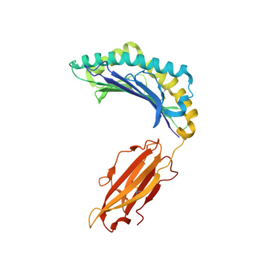
3VFV

crystal structure of HLA B*3508 LPEP-P9Ala, peptide mutant P9-ala


Experimental Data Snapshot

  • Method: X-RAY DIFFRACTION
  • Resolution: 1.55 Å
  • R-Value Free: 0.204 
  • R-Value Work: 0.170 
  • R-Value Observed: 0.172 

Starting Model: experimental
View more details

wwPDB Validation   3D Report Full Report


This is version 1.4 of the entry. See complete history


Literature

The Energetic Basis Underpinning T-cell Receptor Recognition of a Super-bulged Peptide Bound to a Major Histocompatibility Complex Class I Molecule.

Liu, Y.C.Chen, Z.Burrows, S.R.Purcell, A.W.McCluskey, J.Rossjohn, J.Gras, S.

(2012) J Biol Chem 287: 12267-12276

  • DOI: https://doi.org/10.1074/jbc.M112.344689
  • Primary Citation of Related Structures:  
    3VFM, 3VFN, 3VFO, 3VFP, 3VFR, 3VFS, 3VFT, 3VFU, 3VFV, 3VFW

  • PubMed Abstract: 

    Although the major histocompatibility complex class I (MHC-I) molecules typically bind short peptide (p) fragments (8-10 amino acids in length), longer, "bulged" peptides are often be presented by MHC-I. Such bulged pMHC-I complexes represent challenges for T-cell receptor (TCR) ligation, although the general principles underscoring the interaction between TCRs and bulged pMHC-I complexes are unclear. To address this, we have explored the energetic basis of how an immunodominant TCR (termed SB27) binds to a 13-amino acid viral peptide (LPEPLPQGQLTAY) complexed to human leukocyte antigen (HLA) B*3508. Using the crystal structure of the SB27 TCR-HLA B*3508(LPEP) complex as a guide, we undertook a comprehensive alanine-scanning mutagenesis approach at the TCR-pMHC-I interface and examined the effect of the mutations by biophysical (affinity measurements) and cellular approaches (tetramer staining). Although the structural footprint on HLA B*3508 was small, the energetic footprint was even smaller in that only two HLA B*3508 residues were critical for the TCR interaction. Instead, the energetic basis of this TCR-pMHC-I interaction was attributed to peptide-mediated interactions in which the complementarity determining region 3α and germline-encoded complementarity determining region 1β loops of the SB27 TCR played the principal role. Our findings highlight the peptide-centricity of TCR ligation toward a bulged pMHC-I complex.


  • Organizational Affiliation

    Department of Biochemistry and Molecular Biology, Monash University, Clayton, 3800, Australia.


Macromolecules
Find similar proteins by:  (by identity cutoff)  |  3D Structure
Entity ID: 1
MoleculeChains Sequence LengthOrganismDetailsImage
MHC class I antigen276Homo sapiensMutation(s): 0 
Gene Names: HLA-B
UniProt & NIH Common Fund Data Resources
Find proteins for P01889 (Homo sapiens)
Explore P01889 
Go to UniProtKB:  P01889
PHAROS:  P01889
GTEx:  ENSG00000234745 
Entity Groups  
Sequence Clusters30% Identity50% Identity70% Identity90% Identity95% Identity100% Identity
UniProt GroupP01889
Sequence Annotations
Expand
  • Reference Sequence
Find similar proteins by:  (by identity cutoff)  |  3D Structure
Entity ID: 2
MoleculeChains Sequence LengthOrganismDetailsImage
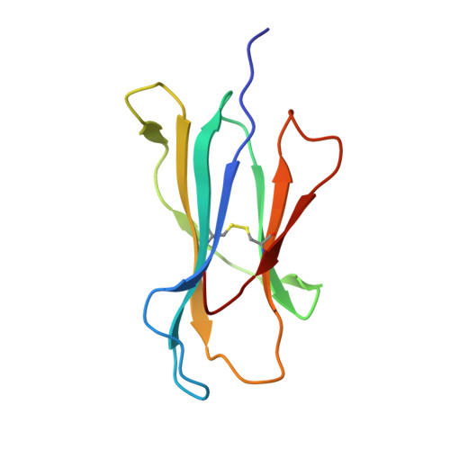
Beta-2-microglobulin100Homo sapiensMutation(s): 0 
Gene Names: B2MCDABP0092HDCMA22P
UniProt & NIH Common Fund Data Resources
Find proteins for P61769 (Homo sapiens)
Explore P61769 
Go to UniProtKB:  P61769
PHAROS:  P61769
GTEx:  ENSG00000166710 
Entity Groups  
Sequence Clusters30% Identity50% Identity70% Identity90% Identity95% Identity100% Identity
UniProt GroupP61769
Sequence Annotations
Expand
  • Reference Sequence

Find similar proteins by:  Sequence   |   3D Structure  

Entity ID: 3
MoleculeChains Sequence LengthOrganismDetailsImage
LPEP peptide from EBV, P9A, LPEPLPQGALTAY13N/AMutation(s): 0 
Entity Groups  
Sequence Clusters30% Identity50% Identity70% Identity90% Identity95% Identity100% Identity
Sequence Annotations
Expand
  • Reference Sequence
Experimental Data & Validation

Experimental Data

  • Method: X-RAY DIFFRACTION
  • Resolution: 1.55 Å
  • R-Value Free: 0.204 
  • R-Value Work: 0.170 
  • R-Value Observed: 0.172 
  • Space Group: P 21 21 21
Unit Cell:
Length ( Å )Angle ( ˚ )
a = 50.8α = 90
b = 81.5β = 90
c = 110.6γ = 90
Software Package:
Software NamePurpose
XSCALEdata scaling
PHASERphasing
PHENIXrefinement
PDB_EXTRACTdata extraction

Structure Validation

View Full Validation Report


Entry History 

Deposition Data

Revision History  (Full details and data files)

  • Version 1.0: 2012-02-22
    Type: Initial release
  • Version 1.1: 2012-03-07
    Changes: Database references
  • Version 1.2: 2012-04-25
    Changes: Database references
  • Version 1.3: 2023-09-13
    Changes: Data collection, Database references, Refinement description
  • Version 1.4: 2024-10-16
    Changes: Structure summary