3UJ1

Crystal structure of the third thioredoxin domain of human ERp46


Experimental Data Snapshot

  • Method: X-RAY DIFFRACTION
  • Resolution: 2.65 Å
  • R-Value Free: 0.259 
  • R-Value Work: 0.229 
  • R-Value Observed: 0.230 

Starting Model: experimental
View more details

wwPDB Validation   3D Report Full Report


This is version 1.3 of the entry. See complete history


Literature

Peptide Binding by Catalytic Domains of the Protein Disulfide Isomerase-Related Protein ERp46.

Funkner, A.Parthier, C.Schutkowski, M.Zerweck, J.Lilie, H.Gyrych, N.Fischer, G.Stubbs, M.T.Ferrari, D.M.

(2013) J Mol Biol 425: 1340-1362

  • DOI: https://doi.org/10.1016/j.jmb.2013.01.029
  • Primary Citation of Related Structures:  
    3UJ1

  • PubMed Abstract: 

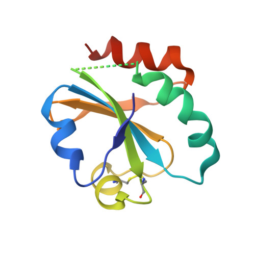
    The protein disulfide isomerase (PDI) family member ERp46/endoPDI/thioredoxin domain-containing protein 5 is preferentially expressed in a limited number of tissues, where it may function as a survival factor for nitrosative stress in vivo. It is involved in insulin production as well as in adiponectin signaling and interacts specifically with the redox-regulatory endoplasmic reticulum proteins endoplasmic oxidoreductin 1α (Ero1α) and peroxiredoxin-4. Here, we show that ERp46, although lacking a PDI-like redox-inactive b'-thioredoxin domain with its hydrophobic substrate binding site, is able to bind to a large pool of peptides containing aromatic and basic residues via all three of its catalytic domains (a(0), a and a'), though the a(0) domain may contain the primary binding site. ERp46, which shows relatively higher activity as a disulfide-reductase than as an oxidase/isomerase in vitro compared to PDI and ERp57, possesses chaperone activity in vivo, a property also shared by the C-terminal a' domain. A crystal structure of the a' domain is also presented, offering a view of possible substrate binding sites within catalytic domains of PDI proteins.


  • Organizational Affiliation

    Max Planck Research Unit for Enzymology of Protein Folding, Weinbergweg 22, 06120 Halle (Saale), Germany.


Macromolecules
Find similar proteins by:  (by identity cutoff)  |  3D Structure
Entity ID: 1
MoleculeChains Sequence LengthOrganismDetailsImage
Thioredoxin domain-containing protein 5110Homo sapiensMutation(s): 0 
Gene Names: TLP46TXNDC5UNQ364/PRO700
UniProt & NIH Common Fund Data Resources
Find proteins for Q8NBS9 (Homo sapiens)
Explore Q8NBS9 
Go to UniProtKB:  Q8NBS9
PHAROS:  Q8NBS9
GTEx:  ENSG00000239264 
Entity Groups  
Sequence Clusters30% Identity50% Identity70% Identity90% Identity95% Identity100% Identity
UniProt GroupQ8NBS9
Sequence Annotations
Expand
  • Reference Sequence
Experimental Data & Validation

Experimental Data

  • Method: X-RAY DIFFRACTION
  • Resolution: 2.65 Å
  • R-Value Free: 0.259 
  • R-Value Work: 0.229 
  • R-Value Observed: 0.230 
  • Space Group: P 32 2 1
Unit Cell:
Length ( Å )Angle ( ˚ )
a = 63.58α = 90
b = 63.58β = 90
c = 68.68γ = 120
Software Package:
Software NamePurpose
MOSFLMdata reduction
SCALAdata scaling
PHASERphasing
REFMACrefinement
PDB_EXTRACTdata extraction
AUTOMARdata collection

Structure Validation

View Full Validation Report


Entry History 

Deposition Data

Revision History  (Full details and data files)

  • Version 1.0: 2012-11-07
    Type: Initial release
  • Version 1.1: 2013-03-06
    Changes: Database references
  • Version 1.2: 2013-04-17
    Changes: Database references
  • Version 1.3: 2023-09-13
    Changes: Data collection, Database references, Refinement description