3TWX

Crystal structure of ARC4 from human Tankyrase 2 in complex with peptide from human FNBP1 (chimeric peptide)


Experimental Data Snapshot

  • Method: X-RAY DIFFRACTION
  • Resolution: 1.80 Å
  • R-Value Free: 0.232 
  • R-Value Work: 0.198 
  • R-Value Observed: 0.200 

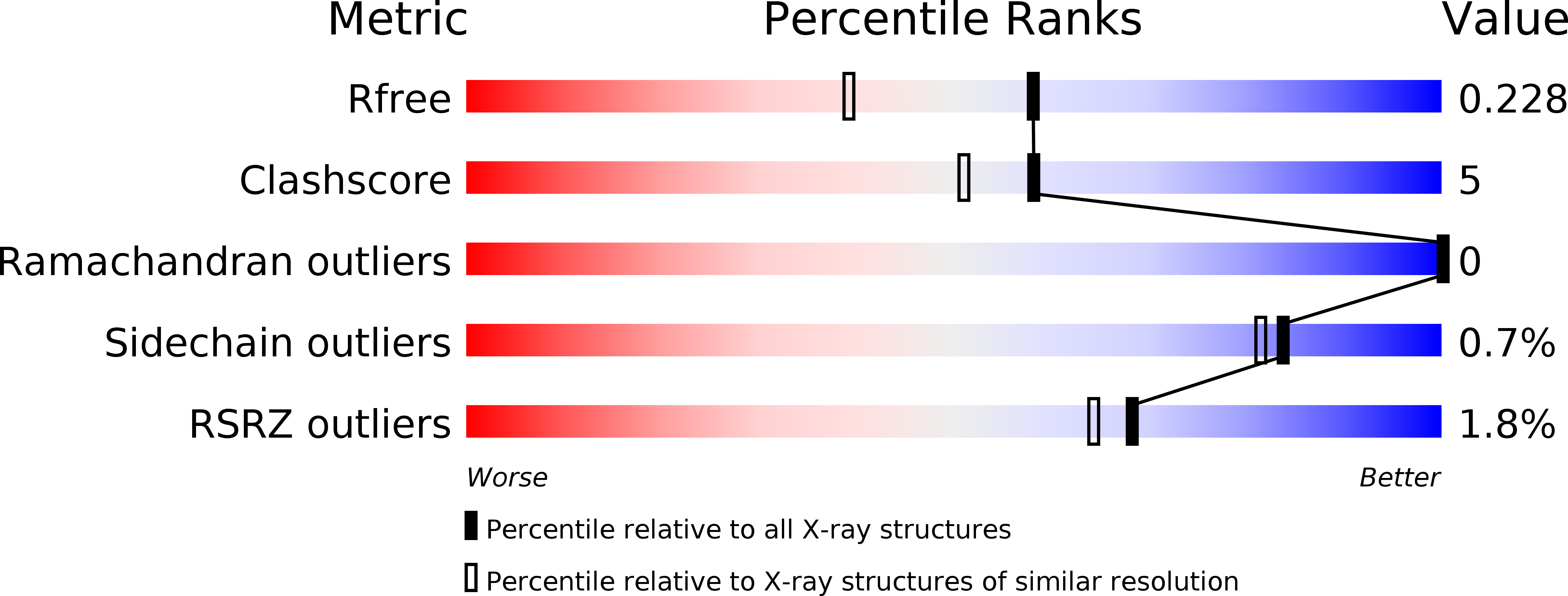
wwPDB Validation   3D Report Full Report


This is version 1.1 of the entry. See complete history


Literature

Structural basis and sequence rules for substrate recognition by tankyrase explain the basis for cherubism disease.

Guettler, S.Larose, J.Petsalaki, E.Gish, G.Scotter, A.Pawson, T.Rottapel, R.Sicheri, F.

(2011) Cell 147: 1340-1354

  • DOI: https://doi.org/10.1016/j.cell.2011.10.046
  • Primary Citation of Related Structures:  
    3TWQ, 3TWR, 3TWS, 3TWT, 3TWU, 3TWV, 3TWW, 3TWX

  • PubMed Abstract: 

    The poly(ADP-ribose)polymerases Tankyrase 1/2 (TNKS/TNKS2) catalyze the covalent linkage of ADP-ribose polymer chains onto target proteins, regulating their ubiquitylation, stability, and function. Dysregulation of substrate recognition by Tankyrases underlies the human disease cherubism. Tankyrases recruit specific motifs (often called RxxPDG "hexapeptides") in their substrates via an N-terminal region of ankyrin repeats. These ankyrin repeats form five domains termed ankyrin repeat clusters (ARCs), each predicted to bind substrate. Here we report crystal structures of a representative ARC of TNKS2 bound to targeting peptides from six substrates. Using a solution-based peptide library screen, we derive a rule-based consensus for Tankyrase substrates common to four functionally conserved ARCs. This 8-residue consensus allows us to rationalize all known Tankyrase substrates and explains the basis for cherubism-causing mutations in the Tankyrase substrate 3BP2. Structural and sequence information allows us to also predict and validate other Tankyrase targets, including Disc1, Striatin, Fat4, RAD54, BCR, and MERIT40.


  • Organizational Affiliation

    Centre for Systems Biology, Samuel Lunenfeld Research Institute, Mount Sinai Hospital, 600 University Avenue, Toronto, Ontario M5G 1X5, Canada.


Macromolecules
Find similar proteins by:  (by identity cutoff)  |  3D Structure
Entity ID: 1
MoleculeChains Sequence LengthOrganismDetailsImage
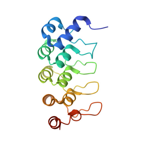
Tankyrase-2
A, B
165Homo sapiensMutation(s): 0 
Gene Names: PARP5BTANK2TNKLTNKS2
EC: 2.4.2.30 (PDB Primary Data), 2.4.2 (UniProt)
UniProt & NIH Common Fund Data Resources
Find proteins for Q9H2K2 (Homo sapiens)
Explore Q9H2K2 
Go to UniProtKB:  Q9H2K2
PHAROS:  Q9H2K2
GTEx:  ENSG00000107854 
Entity Groups  
Sequence Clusters30% Identity50% Identity70% Identity90% Identity95% Identity100% Identity
UniProt GroupQ9H2K2
Sequence Annotations
Expand
  • Reference Sequence

Find similar proteins by:  Sequence   |   3D Structure  

Entity ID: 2
MoleculeChains Sequence LengthOrganismDetailsImage
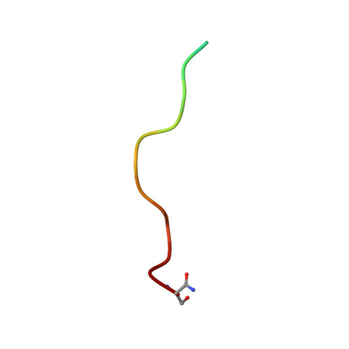
human FNBP1
C, D
16Homo sapiensMutation(s): 0 
Entity Groups  
Sequence Clusters30% Identity50% Identity70% Identity90% Identity95% Identity100% Identity
Sequence Annotations
Expand
  • Reference Sequence
Small Molecules
Modified Residues  1 Unique
IDChains TypeFormula2D DiagramParent
SET
Query on SET
C, D
L-PEPTIDE LINKINGC3 H8 N2 O2SER
Experimental Data & Validation

Experimental Data

  • Method: X-RAY DIFFRACTION
  • Resolution: 1.80 Å
  • R-Value Free: 0.232 
  • R-Value Work: 0.198 
  • R-Value Observed: 0.200 
  • Space Group: P 21 21 21
Unit Cell:
Length ( Å )Angle ( ˚ )
a = 44.196α = 90
b = 74.176β = 90
c = 103.343γ = 90
Software Package:
Software NamePurpose
ADSCdata collection
PHASERphasing
PHENIXrefinement
HKL-2000data reduction
HKL-2000data scaling

Structure Validation

View Full Validation Report


Entry History 

Deposition Data

Revision History  (Full details and data files)

  • Version 1.0: 2011-12-07
    Type: Initial release
  • Version 1.1: 2011-12-28
    Changes: Database references