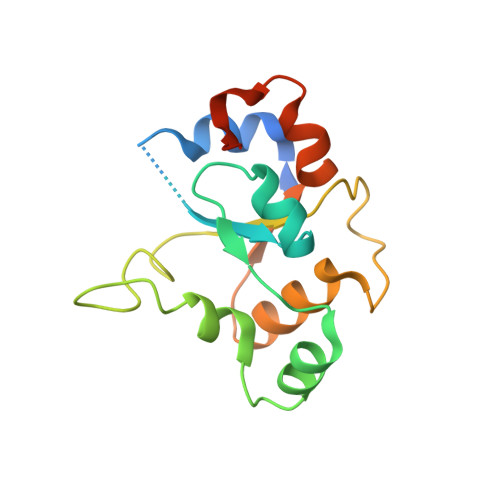
3TG1

Crystal structure of p38alpha in complex with a MAPK docking partner


Experimental Data Snapshot

  • Method: X-RAY DIFFRACTION
  • Resolution: 2.71 Å
  • R-Value Free: 0.260 
  • R-Value Work: 0.216 
  • R-Value Observed: 0.218 

Starting Models: experimental
View more details

wwPDB Validation   3D Report Full Report


This is version 1.1 of the entry. See complete history


Literature

A Distinct Interaction Mode Revealed by the Crystal Structure of the Kinase p38alpha with the MAPK Binding Domain of the Phosphatase MKP5.

Zhang, Y.Y.Wu, J.W.Wang, Z.X.

(2011) Sci Signal 4: ra88-ra88

  • DOI: https://doi.org/10.1126/scisignal.2002241
  • Primary Citation of Related Structures:  
    3TG1, 3TG3

  • PubMed Abstract: 

    The mitogen-activated protein kinase (MAPK) cascades play a pivotal role in a myriad of cellular functions. The specificity and efficiency of MAPK signaling are controlled by docking interactions between MAPKs and their cognate proteins. Many MAPK-interacting partners, including substrates, MAPK kinases, phosphatases, and scaffolding proteins, have linear sequence motifs that mediate the interaction with the common docking site on MAPKs. We report the crystal structure of p38α in complex with the MAPK binding domain (KBD) from MAPK phosphatase 5 (MKP5) at 2.7 Å resolution. In contrast to the well-known docking mode, the KBD binds p38α in a bipartite manner, in which two distinct helical regions of KBD engage the p38α docking site, which is situated on the back of the p38α active site. We also determined the crystal structure of the KBD of MKP7, which closely resembles the MKP5 KBD, suggesting that the mechanism of molecular recognition by the KBD of MKP5 is conserved in the cytoplasmic p38- and c-Jun N-terminal kinase-specific MKP subgroup. This previously unknown binding mode provides new insights into how MAPKs interact with their binding partners to achieve functional specificity.


  • Organizational Affiliation

    Ministry of Education Key Laboratory for Protein Science, School of Life Sciences, Tsinghua University, Beijing 100084, PR China.


Macromolecules
Find similar proteins by:  (by identity cutoff)  |  3D Structure
Entity ID: 1
MoleculeChains Sequence LengthOrganismDetailsImage
Mitogen-activated protein kinase 14380Mus musculusMutation(s): 0 
Gene Names: Mapk14Crk1Csbp1Csbp2
EC: 2.7.11.24
UniProt
Find proteins for P47811 (Mus musculus)
Explore P47811 
Go to UniProtKB:  P47811
Entity Groups  
Sequence Clusters30% Identity50% Identity70% Identity90% Identity95% Identity100% Identity
UniProt GroupP47811
Sequence Annotations
Expand
  • Reference Sequence
Find similar proteins by:  (by identity cutoff)  |  3D Structure
Entity ID: 2
MoleculeChains Sequence LengthOrganismDetailsImage
Dual specificity protein phosphatase 10158Homo sapiensMutation(s): 0 
Gene Names: DUSP10MKP5
EC: 3.1.3.16 (PDB Primary Data), 3.1.3.48 (PDB Primary Data)
UniProt & NIH Common Fund Data Resources
Find proteins for Q9Y6W6 (Homo sapiens)
Explore Q9Y6W6 
Go to UniProtKB:  Q9Y6W6
PHAROS:  Q9Y6W6
GTEx:  ENSG00000143507 
Entity Groups  
Sequence Clusters30% Identity50% Identity70% Identity90% Identity95% Identity100% Identity
UniProt GroupQ9Y6W6
Sequence Annotations
Expand
  • Reference Sequence
Experimental Data & Validation

Experimental Data

  • Method: X-RAY DIFFRACTION
  • Resolution: 2.71 Å
  • R-Value Free: 0.260 
  • R-Value Work: 0.216 
  • R-Value Observed: 0.218 
  • Space Group: P 41 21 2
Unit Cell:
Length ( Å )Angle ( ˚ )
a = 72.428α = 90
b = 72.428β = 90
c = 226.143γ = 90
Software Package:
Software NamePurpose
MAR345dtbdata collection
PHASERphasing
PHENIXrefinement
HKL-2000data reduction
HKL-2000data scaling

Structure Validation

View Full Validation Report


Entry History 

Deposition Data

Revision History  (Full details and data files)

  • Version 1.0: 2012-03-14
    Type: Initial release
  • Version 1.1: 2023-11-01
    Changes: Data collection, Database references, Refinement description