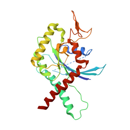
3T5D

Crystal structure of Septin 7 in complex with GDP


Experimental Data Snapshot

  • Method: X-RAY DIFFRACTION
  • Resolution: 3.30 Å
  • R-Value Free: 0.318 
  • R-Value Work: 0.285 
  • R-Value Observed: 0.287 

wwPDB Validation   3D Report Full Report


Ligand Structure Quality Assessment 


This is version 1.1 of the entry. See complete history


Literature

Structural and biochemical properties of Sept7, a unique septin required for filament formation.

Zent, E.Vetter, I.Wittinghofer, A.

(2011) Biol Chem 392: 791-797

  • DOI: https://doi.org/10.1515/BC.2011.082
  • Primary Citation of Related Structures:  
    3T5D

  • PubMed Abstract: 

    Septins constitute a family of conserved guanine nucleotide binding proteins found in a wide range of organisms from fungi to mammals. Members of the family share a canonical G-domain with N- and C-terminal extensions. G-domains assemble into hetero-oligomeric complexes which form non-polarised filaments or rings. Linear filaments are formed between the G-domains using either the guanine nucleotide binding site (G interface) or N- and C-terminal extensions (NC interface). Sept7 is a unique among the 13 human septins in that it occupies the ends of hexameric building blocks which assemble into non-polarised filaments. To gain insight into its particular properties we performed structural and biochemical studies on Sept7. We solved the crystal structure of a Sept7 dimer in the GDP-bound state. The structure and biochemistry of Sept7 provide new insights into the dynamics of the G interface and outline the differences in the properties of Sept7 compared to the members of group 2 septins.


  • Organizational Affiliation

    Structural Biology Group, Max Planck Institute of Molecular Physiology, Dortmund, Germany.


Macromolecules
Find similar proteins by:  (by identity cutoff)  |  3D Structure
Entity ID: 1
MoleculeChains Sequence LengthOrganismDetailsImage
Septin-7A,
B [auth C]
274Homo sapiensMutation(s): 0 
Gene Names: SEPT7CDC10
UniProt & NIH Common Fund Data Resources
Find proteins for Q16181 (Homo sapiens)
Explore Q16181 
Go to UniProtKB:  Q16181
PHAROS:  Q16181
GTEx:  ENSG00000122545 
Entity Groups  
Sequence Clusters30% Identity50% Identity70% Identity90% Identity95% Identity100% Identity
UniProt GroupQ16181
Sequence Annotations
Expand
  • Reference Sequence
Small Molecules
Ligands 1 Unique
IDChains Name / Formula / InChI Key2D Diagram3D Interactions
GDP
Query on GDP

Download Ideal Coordinates CCD File 
C [auth A],
D [auth C]
GUANOSINE-5'-DIPHOSPHATE
C10 H15 N5 O11 P2
QGWNDRXFNXRZMB-UUOKFMHZSA-N
Experimental Data & Validation

Experimental Data

  • Method: X-RAY DIFFRACTION
  • Resolution: 3.30 Å
  • R-Value Free: 0.318 
  • R-Value Work: 0.285 
  • R-Value Observed: 0.287 
  • Space Group: P 61
Unit Cell:
Length ( Å )Angle ( ˚ )
a = 82.363α = 90
b = 82.363β = 90
c = 210.598γ = 120
Software Package:
Software NamePurpose
PROdata collection
MOLREPphasing
REFMACrefinement
XDSdata reduction
XSCALEdata scaling

Structure Validation

View Full Validation Report


Ligand Structure Quality Assessment 


Entry History 

Deposition Data

Revision History  (Full details and data files)

  • Version 1.0: 2011-08-24
    Type: Initial release
  • Version 1.1: 2024-02-28
    Changes: Data collection, Database references, Derived calculations, Refinement description