3SGM

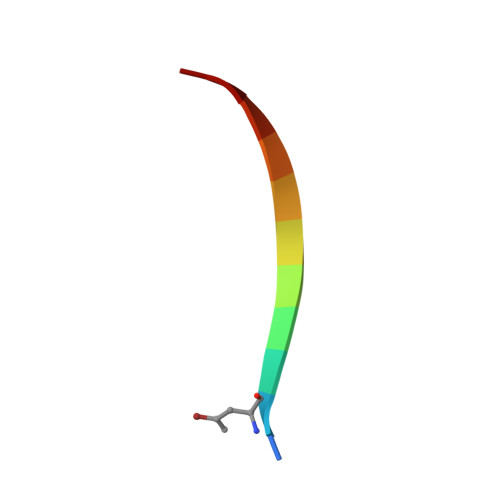
Bromoderivative-2 of amyloid-related segment of alphaB-crystallin residues 90-100


Experimental Data Snapshot

  • Method: X-RAY DIFFRACTION
  • Resolution: 1.70 Å
  • R-Value Free: 0.240 
  • R-Value Work: 0.192 
  • R-Value Observed: 0.194 

wwPDB Validation   3D Report Full Report


This is version 1.0 of the entry. See complete history


Literature

Atomic view of a toxic amyloid small oligomer.

Laganowsky, A.Liu, C.Sawaya, M.R.Whitelegge, J.P.Park, J.Zhao, M.Pensalfini, A.Soriaga, A.B.Landau, M.Teng, P.K.Cascio, D.Glabe, C.Eisenberg, D.

(2012) Science 335: 1228-1231

  • DOI: https://doi.org/10.1126/science.1213151
  • Primary Citation of Related Structures:  
    3SGM, 3SGN, 3SGO, 3SGP, 3SGR, 3SGS

  • PubMed Abstract: 

    Amyloid diseases, including Alzheimer's, Parkinson's, and the prion conditions, are each associated with a particular protein in fibrillar form. These amyloid fibrils were long suspected to be the disease agents, but evidence suggests that smaller, often transient and polymorphic oligomers are the toxic entities. Here, we identify a segment of the amyloid-forming protein αB crystallin, which forms an oligomeric complex exhibiting properties of other amyloid oligomers: β-sheet-rich structure, cytotoxicity, and recognition by an oligomer-specific antibody. The x-ray-derived atomic structure of the oligomer reveals a cylindrical barrel, formed from six antiparallel protein strands, that we term a cylindrin. The cylindrin structure is compatible with a sequence segment from the β-amyloid protein of Alzheimer's disease. Cylindrins offer models for the hitherto elusive structures of amyloid oligomers.


  • Organizational Affiliation

    Department of Biological Chemistry, University of California Los Angeles (UCLA), Howard Hughes Medical Institute (HHMI), Los Angeles, CA 90095, USA.


Macromolecules

Find similar proteins by:  Sequence   |   3D Structure  

Entity ID: 1
MoleculeChains Sequence LengthOrganismDetailsImage
Alpha-crystallin B chain
A, B, C, D
11Homo sapiensMutation(s): 0 
UniProt & NIH Common Fund Data Resources
Find proteins for P02511 (Homo sapiens)
Explore P02511 
Go to UniProtKB:  P02511
PHAROS:  P02511
GTEx:  ENSG00000109846 
Entity Groups  
Sequence Clusters30% Identity50% Identity70% Identity90% Identity95% Identity100% Identity
UniProt GroupP02511
Sequence Annotations
Expand
  • Reference Sequence
Small Molecules
Ligands 1 Unique
IDChains Name / Formula / InChI Key2D Diagram3D Interactions
MPD
Query on MPD

Download Ideal Coordinates CCD File 
E [auth B](4S)-2-METHYL-2,4-PENTANEDIOL
C6 H14 O2
SVTBMSDMJJWYQN-YFKPBYRVSA-N
Modified Residues  1 Unique
IDChains TypeFormula2D DiagramParent
A8E
Query on A8E
A, B, C, D
L-PEPTIDE LINKINGC5 H8 Br N O2VAL
Experimental Data & Validation

Experimental Data

  • Method: X-RAY DIFFRACTION
  • Resolution: 1.70 Å
  • R-Value Free: 0.240 
  • R-Value Work: 0.192 
  • R-Value Observed: 0.194 
  • Space Group: I 21 3
Unit Cell:
Length ( Å )Angle ( ˚ )
a = 65.995α = 90
b = 65.995β = 90
c = 65.995γ = 90
Software Package:
Software NamePurpose
DENZOdata reduction
SCALEPACKdata scaling
SHELXphasing
DMphasing
PHENIXrefinement
PDB_EXTRACTdata extraction
SHELXDphasing

Structure Validation

View Full Validation Report


Entry History 

Deposition Data

Revision History  (Full details and data files)

  • Version 1.0: 2012-03-21
    Type: Initial release