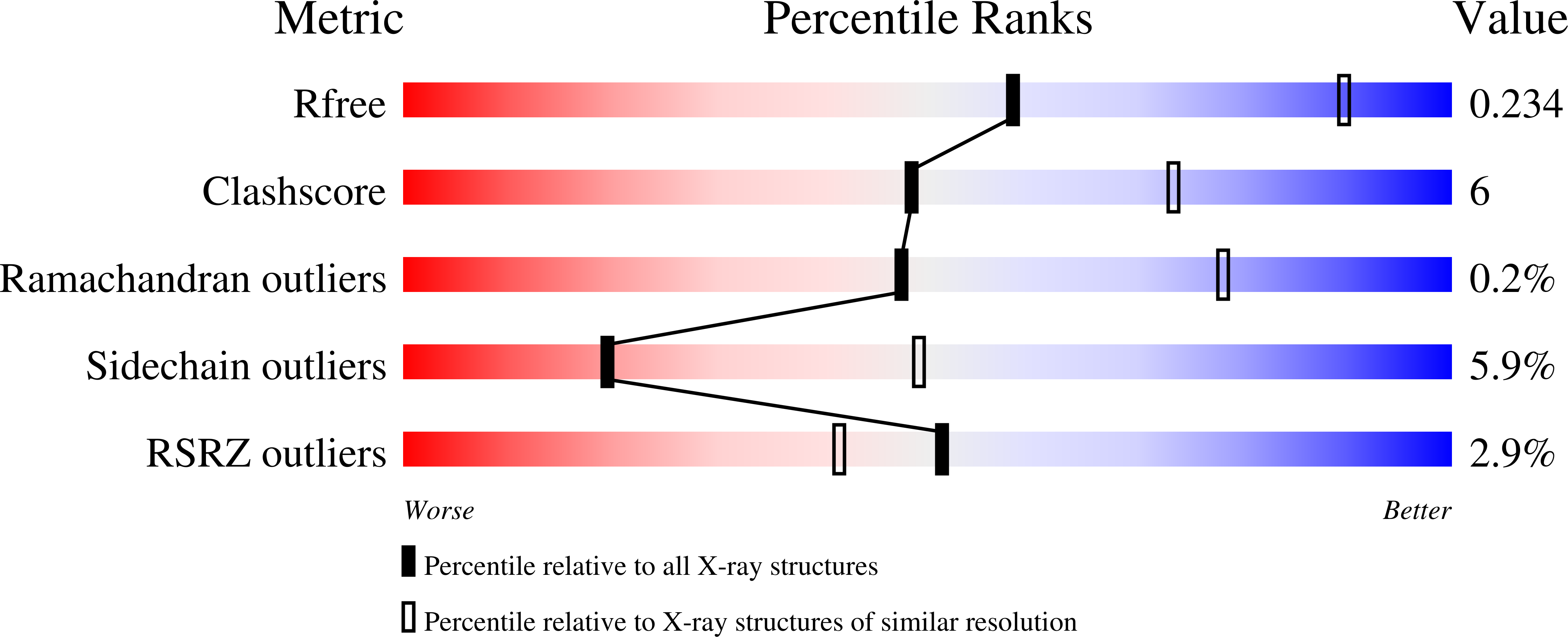
Structure of human lysine methyltransferase Smyd2 reveals insights into the substrate divergence in Smyd proteins
Xu, S., Zhong, C., Zhang, T., Ding, J.(2011) J Mol Cell Biol 3: 293-300
- PubMed: 21724641
- DOI: https://doi.org/10.1093/jmcb/mjr015
- Primary Citation of Related Structures:
3RIB - PubMed Abstract:
The SET- and myeloid-Nervy-DEAF-1 (MYND)-domain containing (Smyd) lysine methyltransferases 1-3 share relatively high sequence similarity but exhibit divergence in the substrate specificity. Here we report the crystal structure of the full-length human Smyd2 in complex with S-adenosyl-L-homocysteine (AdoHcy). Although the Smyd1-3 enzymes are similar in the overall structure, detailed comparisons demonstrate that they differ substantially in the potential substrate-binding site. The binding site of Smyd3 consists mainly of a deep and narrow pocket, while those of Smyd1 and Smyd2 consist of a comparable pocket and a long groove. In addition, Smyd2, which has lysine methyltransferase activity on histone H3-lysine 36, exhibits substantial differences in the wall of the substrate-binding pocket compared with those of Smyd1 and Smyd3 which have activity specifically on histone H3-lysine 4. The differences in the substrate-binding site might account for the observed divergence in the specificity and methylation state of the substrates. Further modeling study of Smyd2 in complex with a p53 peptide indicates that mono-methylation of p53-Lys(372) might result in steric conflict of the methyl group with the surrounding residues of Smyd2, providing a structural explanation for the inhibitory effect of the SET7/9-mediated mono-methylation of p53-Lys(372) on the Smyd2-mediated methylation of p53-Lys(370).
Organizational Affiliation:
State Key Laboratory of Molecular Biology and Research Center for Structural Biology, Institute of Biochemistry and Cell Biology, Shanghai Institutes for Biological Sciences, Chinese Academy of Sciences, China.


