3R7N

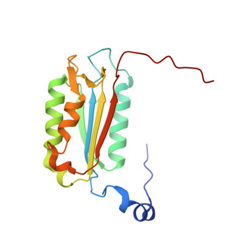
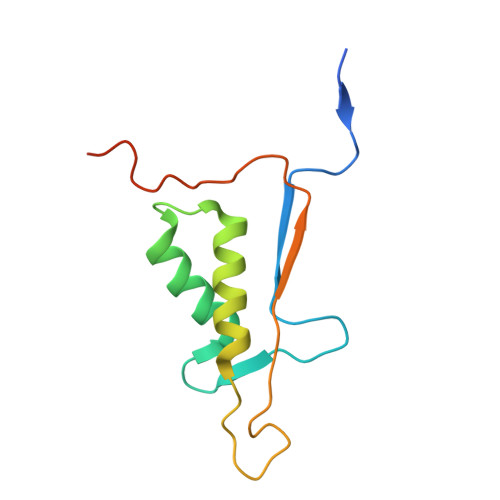
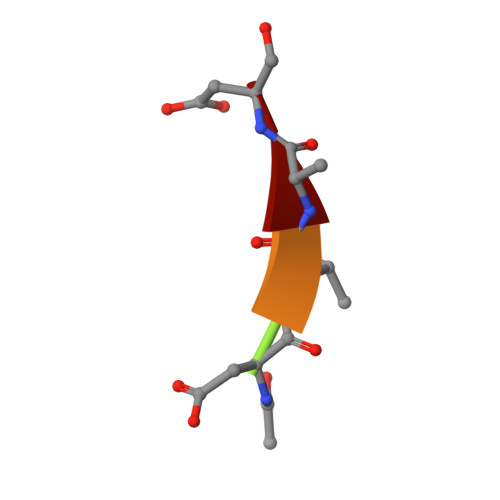
Caspase-2 bound with two copies of Ac-DVAD-CHO


Experimental Data Snapshot

  • Method: X-RAY DIFFRACTION
  • Resolution: 2.33 Å
  • R-Value Free: 0.227 
  • R-Value Work: 0.172 
  • R-Value Observed: 0.175 

wwPDB Validation   3D Report Full Report


This is version 1.2 of the entry. See complete history


Literature

Structural and enzymatic insights into caspase-2 protein substrate recognition and catalysis.

Tang, Y.Wells, J.A.Arkin, M.R.

(2011) J Biol Chem 286: 34147-34154

  • DOI: https://doi.org/10.1074/jbc.M111.247627
  • Primary Citation of Related Structures:  
    3R5J, 3R6G, 3R6L, 3R7B, 3R7N, 3R7S

  • PubMed Abstract: 

    Caspase-2, the most evolutionarily conserved member in the human caspase family, may play important roles in stress-induced apoptosis, cell cycle regulation, and tumor suppression. In biochemical assays, caspase-2 uniquely prefers a pentapeptide (such as VDVAD) rather than a tetrapeptide, as required for efficient cleavage by other caspases. We investigated the molecular basis for pentapeptide specificity using peptide analog inhibitors and substrates that vary at the P5 position. We determined the crystal structures of apo caspase-2, caspase-2 in complex with peptide inhibitors VDVAD-CHO, ADVAD-CHO, and DVAD-CHO, and a T380A mutant of caspase-2 in complex with VDVAD-CHO. Two residues, Thr-380 and Tyr-420, are identified to be critical for the P5 residue recognition; mutation of the two residues reduces the catalytic efficiency by about 4- and 40-fold, respectively. The structures also provide a series of snapshots of caspase-2 in different catalytic states, shedding light on the mechanism of capase-2 activation, substrate binding, and catalysis. By comparing the apo and inhibited caspase-2 structures, we propose that the disruption of a non-conserved salt bridge between Glu-217 and the invariant Arg-378 is important for the activation of caspase-2. These findings broaden our understanding of caspase-2 substrate specificity and catalysis.


  • Organizational Affiliation

    Department of Pharmaceutical Chemistry, Small Molecule Discovery Center, University of California, San Francisco, California 94158, USA.


Macromolecules
Find similar proteins by:  (by identity cutoff)  |  3D Structure
Entity ID: 1
MoleculeChains Sequence LengthOrganismDetailsImage
Caspase-2 subunit p18
A, C
160Homo sapiensMutation(s): 0 
Gene Names: CASP2ICH1NEDD2
EC: 3.4.22 (PDB Primary Data), 3.4.22.55 (UniProt)
UniProt & NIH Common Fund Data Resources
Find proteins for P42575 (Homo sapiens)
Explore P42575 
Go to UniProtKB:  P42575
PHAROS:  P42575
GTEx:  ENSG00000106144 
Entity Groups  
Sequence Clusters30% Identity50% Identity70% Identity90% Identity95% Identity100% Identity
UniProt GroupP42575
Sequence Annotations
Expand
  • Reference Sequence
Find similar proteins by:  (by identity cutoff)  |  3D Structure
Entity ID: 2
MoleculeChains Sequence LengthOrganismDetailsImage
Caspase-2 subunit p12
B, D
112Homo sapiensMutation(s): 0 
Gene Names: CASP2ICH1NEDD2
EC: 3.4.22 (PDB Primary Data), 3.4.22.55 (UniProt)
UniProt & NIH Common Fund Data Resources
Find proteins for P42575 (Homo sapiens)
Explore P42575 
Go to UniProtKB:  P42575
PHAROS:  P42575
GTEx:  ENSG00000106144 
Entity Groups  
Sequence Clusters30% Identity50% Identity70% Identity90% Identity95% Identity100% Identity
UniProt GroupP42575
Sequence Annotations
Expand
  • Reference Sequence

Find similar proteins by:  Sequence   |   3D Structure  

Entity ID: 3
MoleculeChains Sequence LengthOrganismDetailsImage
Peptide Inhibitor (ACE)DVAD-CHOE [auth F],
F [auth E]
5N/AMutation(s): 0 
Sequence Annotations
Expand
  • Reference Sequence
Small Molecules
Modified Residues  1 Unique
IDChains TypeFormula2D DiagramParent
ASA
Query on ASA
E [auth F],
F [auth E]
L-PEPTIDE LINKINGC4 H7 N O3ASP
Biologically Interesting Molecules (External Reference) 1 Unique
Experimental Data & Validation

Experimental Data

  • Method: X-RAY DIFFRACTION
  • Resolution: 2.33 Å
  • R-Value Free: 0.227 
  • R-Value Work: 0.172 
  • R-Value Observed: 0.175 
  • Space Group: P 21 21 21
Unit Cell:
Length ( Å )Angle ( ˚ )
a = 62.876α = 90
b = 96.856β = 90
c = 98.142γ = 90
Software Package:
Software NamePurpose
Blu-Icedata collection
MOLREPphasing
REFMACrefinement
HKL-2000data reduction
HKL-2000data scaling

Structure Validation

View Full Validation Report


Entry History 

Deposition Data

Revision History  (Full details and data files)

  • Version 1.0: 2011-07-27
    Type: Initial release
  • Version 1.1: 2011-10-19
    Changes: Database references
  • Version 1.2: 2012-12-12
    Changes: Other