3Q4Z

Structure of unphosphorylated PAK1 kinase domain


Experimental Data Snapshot

  • Method: X-RAY DIFFRACTION
  • Resolution: 1.89 Å
  • R-Value Free: 0.231 
  • R-Value Work: 0.184 
  • R-Value Observed: 0.186 

Starting Model: experimental
View more details

wwPDB Validation   3D Report Full Report


Ligand Structure Quality Assessment 


This is version 1.2 of the entry. See complete history


Literature

Structural insights into the autoactivation mechanism of p21-activated protein kinase

Wang, J.Wu, J.-W.Wang, Z.-X.

(2011) Structure 19: 1752-1761

  • DOI: https://doi.org/10.1016/j.str.2011.10.013
  • Primary Citation of Related Structures:  
    3Q4Z, 3Q52, 3Q53

  • PubMed Abstract: 

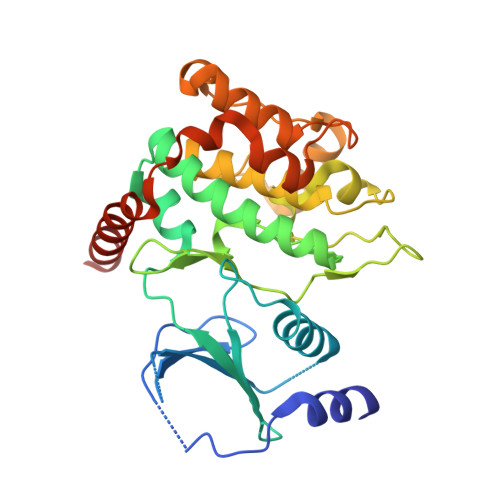
    p21-activated kinases (PAKs) play an important role in diverse cellular processes. Full activation of PAKs requires autophosphorylation of a critical threonine/serine located in the activation loop of the kinase domain. Here we report crystal structures of the phosphorylated and unphosphorylated PAK1 kinase domain. The phosphorylated PAK1 kinase domain has a conformation typical of all active protein kinases. Interestingly, the structure of the unphosphorylated PAK1 kinase domain reveals an unusual dimeric arrangement expected in an authentic enzyme-substrate complex, in which the activation loop of the putative "substrate" is projected into the active site of the "enzyme." The enzyme is bound to AMP-PNP and has an active conformation, whereas the substrate is empty and adopts an inactive conformation. Thus, the structure of the asymmetric homodimer mimics a trans-autophosphorylation complex, and suggests that unphosphorylated PAK1 could dynamically adopt both the active and inactive conformations in solution.


  • Organizational Affiliation

    MOE Key Laboratory for Protein Science, School of Life Sciences, Tsinghua University, Beijing 100084, China.


Macromolecules
Find similar proteins by:  (by identity cutoff)  |  3D Structure
Entity ID: 1
MoleculeChains Sequence LengthOrganismDetailsImage
Serine/threonine-protein kinase PAK 1
A, B
306Homo sapiensMutation(s): 4 
Gene Names: PAK1
EC: 2.7.11.1
UniProt & NIH Common Fund Data Resources
Find proteins for Q13153 (Homo sapiens)
Explore Q13153 
Go to UniProtKB:  Q13153
GTEx:  ENSG00000149269 
Entity Groups  
Sequence Clusters30% Identity50% Identity70% Identity90% Identity95% Identity100% Identity
UniProt GroupQ13153
Sequence Annotations
Expand
  • Reference Sequence
Small Molecules
Ligands 2 Unique
IDChains Name / Formula / InChI Key2D Diagram3D Interactions
ANP
Query on ANP

Download Ideal Coordinates CCD File 
D [auth A]PHOSPHOAMINOPHOSPHONIC ACID-ADENYLATE ESTER
C10 H17 N6 O12 P3
PVKSNHVPLWYQGJ-KQYNXXCUSA-N
MG
Query on MG

Download Ideal Coordinates CCD File 
C [auth A]MAGNESIUM ION
Mg
JLVVSXFLKOJNIY-UHFFFAOYSA-N
Experimental Data & Validation

Experimental Data

  • Method: X-RAY DIFFRACTION
  • Resolution: 1.89 Å
  • R-Value Free: 0.231 
  • R-Value Work: 0.184 
  • R-Value Observed: 0.186 
  • Space Group: P 1 21 1
Unit Cell:
Length ( Å )Angle ( ˚ )
a = 62.988α = 90
b = 79.996β = 107.51
c = 65.664γ = 90
Software Package:
Software NamePurpose
MAR345dtbdata collection
PHASERphasing
PHENIXrefinement
HKL-2000data reduction
HKL-2000data scaling

Structure Validation

View Full Validation Report


Ligand Structure Quality Assessment 


Entry History 

Deposition Data

Revision History  (Full details and data files)

  • Version 1.0: 2011-12-21
    Type: Initial release
  • Version 1.1: 2012-04-11
    Changes: Database references
  • Version 1.2: 2023-11-01
    Changes: Data collection, Database references, Derived calculations, Refinement description