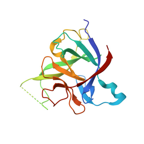
Engineering encodable lanthanide-binding tags into loop regions of proteins.
Barthelmes, K., Reynolds, A.M., Peisach, E., Jonker, H.R., DeNunzio, N.J., Allen, K.N., Imperiali, B., Schwalbe, H.(2011) J Am Chem Soc 133: 808-819
- PubMed: 21182275
- DOI: https://doi.org/10.1021/ja104983t
- Primary Citation of Related Structures:
3LTQ, 3POK - PubMed Abstract:
Lanthanide-binding tags (LBTs) are valuable tools for investigation of protein structure, function, and dynamics by NMR spectroscopy, X-ray crystallography, and luminescence studies. We have inserted LBTs into three different loop positions (denoted L, R, and S) of the model protein interleukin-1β (IL1β) and varied the length of the spacer between the LBT and the protein (denoted 1−3). Luminescence studies demonstrate that all nine constructs bind Tb3+ tightly in the low nanomolar range. No significant change in the fusion protein occurs from insertion of the LBT, as shown by two X-ray crystallographic structures of the IL1β-S1 and IL1β-L3 constructs and for the remaining constructs by comparing the 1H−15N heteronuclear single-quantum coherence NMR spectra with that of the wild-type IL1β. Additionally, binding of LBT-loop IL1β proteins to their native binding partner in vitro remains unaltered. X-ray crystallographic phasing was successful using only the signal from the bound lanthanide. Large residual dipolar couplings (RDCs) could be determined by NMR spectroscopy for all LBT-loop constructs and revealed that the LBT-2 series were rigidly incorporated into the interleukin-1β structure. The paramagnetic NMR spectra of loop-LBT mutant IL1β-R2 were assigned and the Δχ tensor components were calculated on the basis of RDCs and pseudocontact shifts. A structural model of the IL1β-R2 construct was calculated using the paramagnetic restraints. The current data provide support that encodable LBTs serve as versatile biophysical tags when inserted into loop regions of proteins of known structure or predicted via homology modeling.
Organizational Affiliation:
Institute for Organic Chemistry and Chemical Biology, Center for Biomolecular Magnetic Resonance, Johann Wolfgang Goethe-University of Frankfurt, Max-von-Laue-Strasse 7, 60438 Frankfurt, Germany.


