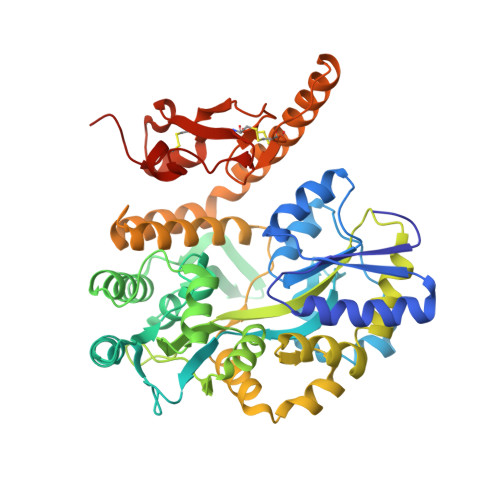
3N94

Crystal structure of human pituitary adenylate cyclase 1 Receptor-short N-terminal extracellular domain


Experimental Data Snapshot

  • Method: X-RAY DIFFRACTION
  • Resolution: 1.80 Å
  • R-Value Free: 0.216 
  • R-Value Work: 0.176 
  • R-Value Observed: 0.178 

Starting Model: experimental
View more details

wwPDB Validation   3D Report Full Report


This is version 2.2 of the entry. See complete history


Literature

Crystal Structure of the PAC1R Extracellular Domain Unifies a Consensus Fold for Hormone Recognition by Class B G-Protein Coupled Receptors.

Kumar, S.Pioszak, A.Zhang, C.Swaminathan, K.Xu, H.E.

(2011) PLoS One 6: e19682-e19682

  • DOI: https://doi.org/10.1371/journal.pone.0019682
  • Primary Citation of Related Structures:  
    3N94

  • PubMed Abstract: 

    Pituitary adenylate cyclase activating polypeptide (PACAP) is a member of the PACAP/glucagon family of peptide hormones, which controls many physiological functions in the immune, nervous, endocrine, and muscular systems. It activates adenylate cyclase by binding to its receptor, PAC1R, a member of class B G-protein coupled receptors (GPCR). Crystal structures of a number of Class B GPCR extracellular domains (ECD) bound to their respective peptide hormones have revealed a consensus mechanism of hormone binding. However, the mechanism of how PACAP binds to its receptor remains controversial as an NMR structure of the PAC1R ECD/PACAP complex reveals a different topology of the ECD and a distinct mode of ligand recognition. Here we report a 1.9 Å crystal structure of the PAC1R ECD, which adopts the same fold as commonly observed for other members of Class B GPCR. Binding studies and cell-based assays with alanine-scanned peptides and mutated receptor support a model that PAC1R uses the same conserved fold of Class B GPCR ECD for PACAP binding, thus unifying the consensus mechanism of hormone binding for this family of receptors.


  • Organizational Affiliation

    Laboratory of Structural Sciences, Van Andel Research Institute, Grand Rapids, Michigan, United States of America.


Macromolecules
Find similar proteins by:  (by identity cutoff)  |  3D Structure
Entity ID: 1
MoleculeChains Sequence LengthOrganismDetailsImage
Fusion protein of Maltose-binding periplasmic protein and pituitary adenylate cyclase 1 Receptor-short475Escherichia coli K-12Homo sapiens
This entity is chimeric
Mutation(s): 0 
Gene Names: malEb4034JW3994ADCYAP1R1
UniProt & NIH Common Fund Data Resources
Find proteins for P41586 (Homo sapiens)
Explore P41586 
Go to UniProtKB:  P41586
PHAROS:  P41586
GTEx:  ENSG00000078549 
Find proteins for P0AEX9 (Escherichia coli (strain K12))
Explore P0AEX9 
Go to UniProtKB:  P0AEX9
Entity Groups  
Sequence Clusters30% Identity50% Identity70% Identity90% Identity95% Identity100% Identity
UniProt GroupsP0AEX9P41586
Sequence Annotations
Expand
  • Reference Sequence
Oligosaccharides

Help

Entity ID: 2
MoleculeChains Length2D Diagram Glycosylation3D Interactions
alpha-D-glucopyranose-(1-4)-alpha-D-glucopyranose
B
2N/A
Glycosylation Resources
GlyTouCan:  G07411ON
GlyCosmos:  G07411ON
Small Molecules
Ligands 1 Unique
IDChains Name / Formula / InChI Key2D Diagram3D Interactions
SO4
Query on SO4

Download Ideal Coordinates CCD File 
C [auth A]SULFATE ION
O4 S
QAOWNCQODCNURD-UHFFFAOYSA-L
Biologically Interesting Molecules (External Reference) 1 Unique
Experimental Data & Validation

Experimental Data

  • Method: X-RAY DIFFRACTION
  • Resolution: 1.80 Å
  • R-Value Free: 0.216 
  • R-Value Work: 0.176 
  • R-Value Observed: 0.178 
  • Space Group: P 21 21 21
Unit Cell:
Length ( Å )Angle ( ˚ )
a = 45.987α = 90
b = 92.151β = 90
c = 105.418γ = 90
Software Package:
Software NamePurpose
REFMACrefinement
PDB_EXTRACTdata extraction
MAR345dtbdata collection
HKL-2000data reduction
HKL-2000data scaling
PHASERphasing

Structure Validation

View Full Validation Report


Entry History 

Deposition Data

Revision History  (Full details and data files)

  • Version 1.0: 2011-06-08
    Type: Initial release
  • Version 1.1: 2011-07-13
    Changes: Version format compliance
  • Version 1.2: 2017-07-26
    Changes: Refinement description, Source and taxonomy
  • Version 2.0: 2020-07-29
    Type: Remediation
    Reason: Carbohydrate remediation
    Changes: Atomic model, Data collection, Database references, Derived calculations, Non-polymer description, Structure summary
  • Version 2.1: 2023-09-06
    Changes: Data collection, Database references, Refinement description, Structure summary
  • Version 2.2: 2024-10-16
    Changes: Structure summary