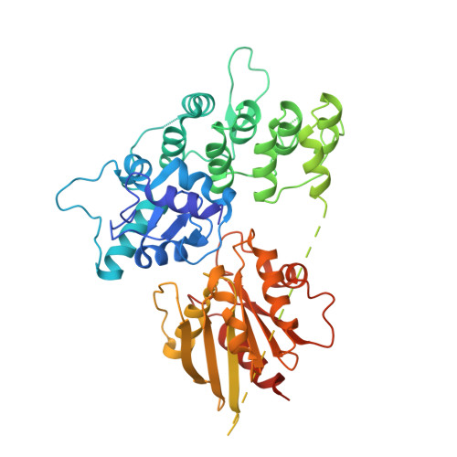
3LVR

The crystal structure of ASAP3 in complex with Arf6 in transition state soaked with Calcium


Experimental Data Snapshot

  • Method: X-RAY DIFFRACTION
  • Resolution: 3.38 Å
  • R-Value Free: 0.281 
  • R-Value Work: 0.207 
  • R-Value Observed: 0.211 

Starting Model: experimental
View more details

wwPDB Validation   3D Report Full Report


Ligand Structure Quality Assessment 


This is version 1.4 of the entry. See complete history


Literature

The structure of an Arf-ArfGAP complex reveals a Ca2+ regulatory mechanism

Ismail, S.A.Vetter, I.R.Sot, B.Wittinghofer, A.

(2010) Cell 141: 812-821

  • DOI: https://doi.org/10.1016/j.cell.2010.03.051
  • Primary Citation of Related Structures:  
    3LVQ, 3LVR

  • PubMed Abstract: 

    Arfs are small G proteins that have a key role in vesicle trafficking and cytoskeletal remodeling. ArfGAP proteins stimulate Arf intrinsic GTP hydrolysis by a mechanism that is still unresolved. Using a fusion construct we solved the structure of the ArfGAP ASAP3 in complex with Arf6 in the transition state. This structure clarifies the ArfGAP catalytic mechanism and shows a glutamine((Arf6)) and an arginine finger((ASAP3)) as the important catalytic residues. Unexpectedly the structure shows a calcium ion, liganded by both proteins in the complex interface, stabilizing the interaction and orienting the catalytic machinery. Calcium stimulates the GAP activity of ASAPs, but not other members of the ArfGAP family. This type of regulation is unique for GAPs and any other calcium-regulated processes and hints at a crosstalk between Ca(2+) and Arf signaling.


  • Organizational Affiliation

    Department of Structural Biology, Max-Planck-Institute für Molekulare Physiologie, Dortmund 44227, Germany.


Macromolecules
Find similar proteins by:  (by identity cutoff)  |  3D Structure
Entity ID: 1
MoleculeChains Sequence LengthOrganismDetailsImage
Arf-GAP with SH3 domain, ANK repeat and PH domain-containing protein 3, ADP-ribosylation factor 6A [auth E]497Homo sapienssynthetic constructMutation(s): 0 
Gene Names: ASAP3ARF6
EC: 3.6.5.2
UniProt & NIH Common Fund Data Resources
Find proteins for P62330 (Homo sapiens)
Explore P62330 
Go to UniProtKB:  P62330
PHAROS:  P62330
GTEx:  ENSG00000165527 
Find proteins for Q8TDY4 (Homo sapiens)
Explore Q8TDY4 
Go to UniProtKB:  Q8TDY4
PHAROS:  Q8TDY4
GTEx:  ENSG00000088280 
Entity Groups  
Sequence Clusters30% Identity50% Identity70% Identity90% Identity95% Identity100% Identity
UniProt GroupsP62330Q8TDY4
Sequence Annotations
Expand
  • Reference Sequence
Experimental Data & Validation

Experimental Data

  • Method: X-RAY DIFFRACTION
  • Resolution: 3.38 Å
  • R-Value Free: 0.281 
  • R-Value Work: 0.207 
  • R-Value Observed: 0.211 
  • Space Group: P 62
Unit Cell:
Length ( Å )Angle ( ˚ )
a = 146.45α = 90
b = 146.45β = 90
c = 49.62γ = 120
Software Package:
Software NamePurpose
ProDCdata collection
MOLREPphasing
REFMACrefinement
XDSdata reduction
XSCALEdata scaling

Structure Validation

View Full Validation Report


Ligand Structure Quality Assessment 


Entry History 

Deposition Data

Revision History  (Full details and data files)

  • Version 1.0: 2010-06-09
    Type: Initial release
  • Version 1.1: 2011-07-13
    Changes: Version format compliance
  • Version 1.2: 2017-08-09
    Changes: Refinement description, Source and taxonomy
  • Version 1.3: 2017-11-08
    Changes: Refinement description
  • Version 1.4: 2023-11-01
    Changes: Data collection, Database references, Derived calculations, Refinement description