3LC8

Crystal structure of the cytoplasmic tail of (pro)renin receptor as a MBP fusion (Maltose-free form)


Experimental Data Snapshot

  • Method: X-RAY DIFFRACTION
  • Resolution: 2.00 Å
  • R-Value Free: 0.257 
  • R-Value Work: 0.195 
  • R-Value Observed: 0.197 

Starting Model: experimental
View more details

wwPDB Validation   3D Report Full Report


This is version 1.3 of the entry. See complete history


Literature

Structural analysis of the intracellular domain of (pro)renin receptor fused to maltose-binding protein.

Zhang, Y.Gao, X.Michael Garavito, R.

(2011) Biochem Biophys Res Commun 407: 674-679

  • DOI: https://doi.org/10.1016/j.bbrc.2011.03.074
  • Primary Citation of Related Structures:  
    3LBS, 3LC8

  • PubMed Abstract: 

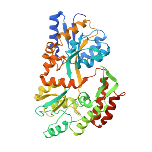
    The (pro)renin receptor (PRR) is an important component of the renin-angiotensin system (RAS), which regulates blood pressure and cardiovascular function. The integral membrane protein PRR contains a large extracellular domain (∼310 amino acids), a single transmembrane domain (∼20 amino acids) and an intracellular domain (∼19 amino acids). Although short, the intracellular (IC) domain of the PRR has functionally important roles in a number of signal transduction pathways activated by (pro)renin binding. Meanwhile, together with the transmembrane domain and a small portion of the extracellular domain (∼30 amino acids), the IC domain is also involved in assembly of V(0) portion of the vacuolar proton-translocating ATPase (V-ATPase). To better understand structural and multifunctional roles of the PRR-IC, we report the crystal structure of the PRR-IC domain as maltose-binding protein (MBP) fusion proteins at 2.0Å (maltose-free) and 2.15Å (maltose-bound). In the two separate crystal forms having significantly different unit-cell dimensions and molecular packing, MBP-PRR-IC fusion protein was found to be a dimer, which is different with the natural monomer of native MBP. The PRR-IC domain appears as a relatively flexible loop and is responsible for the dimerization of MBP fusion protein. Residues in the PRR-IC domain, particularly two tyrosines, dominate the intermonomer interactions, suggesting a role for the PRR-IC domain in protein oligomerization.


  • Organizational Affiliation

    Department of Biochemistry and Molecular Biology, Michigan State University, East Lansing, MI 48824-1319, USA.


Macromolecules
Find similar proteins by:  (by identity cutoff)  |  3D Structure
Entity ID: 1
MoleculeChains Sequence LengthOrganismDetailsImage
Maltose-binding periplasmic protein, Renin receptor
A, B
384Escherichia coliHomo sapiens
This entity is chimeric
Mutation(s): 0 
Gene Names: b4034JW3994malEATP6AP2ATP6IP2CAPERELDF10HT028MSTP009PSEC0072
UniProt & NIH Common Fund Data Resources
Find proteins for P0AEX9 (Escherichia coli (strain K12))
Explore P0AEX9 
Go to UniProtKB:  P0AEX9
Find proteins for O75787 (Homo sapiens)
Explore O75787 
Go to UniProtKB:  O75787
GTEx:  ENSG00000182220 
Entity Groups  
Sequence Clusters30% Identity50% Identity70% Identity90% Identity95% Identity100% Identity
UniProt GroupsP0AEX9O75787
Sequence Annotations
Expand
  • Reference Sequence
Experimental Data & Validation

Experimental Data

  • Method: X-RAY DIFFRACTION
  • Resolution: 2.00 Å
  • R-Value Free: 0.257 
  • R-Value Work: 0.195 
  • R-Value Observed: 0.197 
  • Space Group: P 21 21 21
Unit Cell:
Length ( Å )Angle ( ˚ )
a = 47.78α = 90
b = 112.7β = 90
c = 175.11γ = 90
Software Package:
Software NamePurpose
HKL-2000data collection
AMoREphasing
REFMACrefinement
HKL-2000data reduction
HKL-2000data scaling

Structure Validation

View Full Validation Report


Entry History 

Deposition Data

Revision History  (Full details and data files)

  • Version 1.0: 2011-02-16
    Type: Initial release
  • Version 1.1: 2011-07-13
    Changes: Version format compliance
  • Version 1.2: 2017-08-09
    Changes: Refinement description, Source and taxonomy
  • Version 1.3: 2023-09-06
    Changes: Data collection, Database references, Derived calculations, Refinement description