3K0K

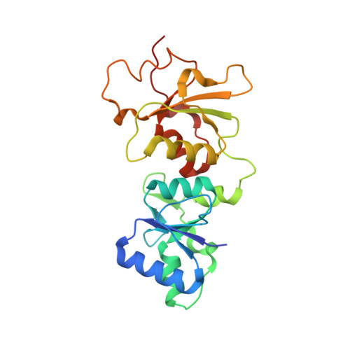
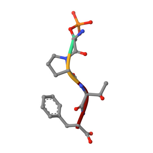
Crystal Structure of BRCA1 BRCT in complex with a minimal recognition tetrapeptide with a free carboxy C-terminus.


Experimental Data Snapshot

  • Method: X-RAY DIFFRACTION
  • Resolution: 2.70 Å
  • R-Value Free: 0.299 
  • R-Value Work: 0.259 
  • R-Value Observed: 0.261 

wwPDB Validation   3D Report Full Report


This is version 1.2 of the entry. See complete history


Literature

Comparison of the Structures and Peptide Binding Specificities of the BRCT Domains of MDC1 and BRCA1

Campbell, S.J.Edwards, R.A.Glover, J.N.

(2010) Structure 18: 167-176

  • DOI: https://doi.org/10.1016/j.str.2009.12.008
  • Primary Citation of Related Structures:  
    3K05, 3K0H, 3K0K, 3K15, 3K16

  • PubMed Abstract: 

    The tandem BRCT domains of BRCA1 and MDC1 facilitate protein signaling at DNA damage foci through specific interactions with serine-phosphorylated protein partners. The MDC1 BRCT binds pSer-Gln-Glu-Tyr-COO(-) at the C terminus of the histone variant gammaH2AX via direct recognition of the C-terminal carboxylate, while BRCA1 recognizes pSer-X-X-Phe motifs either at C-terminal or internal sites within target proteins. Using fluorescence polarization binding assays, we show that while both BRCTs prefer a free main chain carboxylate at the +3 position, this preference is much more pronounced in MDC1. Crystal structures of BRCA1 and MDC1 bound to tetrapeptide substrates reveal differences in the environment of conserved arginines (Arg1699 in BRCA1 and Arg1933 in MDC1) that determine the relative affinity for peptides with -COO(-) versus -CO-NH(2) termini. A mutation in MDC1 that induces a more BRCA1-like conformation relaxes the binding specificity, allowing the mutant to bind phosphopeptides lacking a -COO(-) terminus.


  • Organizational Affiliation

    Department of Biochemistry, University of Alberta, Edmonton, AB T6G 2H7, Canada.


Macromolecules
Find similar proteins by:  (by identity cutoff)  |  3D Structure
Entity ID: 1
MoleculeChains Sequence LengthOrganismDetailsImage
Breast cancer type 1 susceptibility protein215Homo sapiensMutation(s): 0 
Gene Names: BRCA1RNF53
EC: 2.3.2.27
UniProt & NIH Common Fund Data Resources
Find proteins for P38398 (Homo sapiens)
Explore P38398 
Go to UniProtKB:  P38398
PHAROS:  P38398
GTEx:  ENSG00000012048 
Entity Groups  
Sequence Clusters30% Identity50% Identity70% Identity90% Identity95% Identity100% Identity
UniProt GroupP38398
Sequence Annotations
Expand
  • Reference Sequence

Find similar proteins by:  Sequence   |   3D Structure  

Entity ID: 2
MoleculeChains Sequence LengthOrganismDetailsImage
phospho peptide pSPTF-COOH4Homo sapiensMutation(s): 0 
Sequence Annotations
Expand
  • Reference Sequence
Small Molecules
Ligands 2 Unique
IDChains Name / Formula / InChI Key2D Diagram3D Interactions
NI
Query on NI

Download Ideal Coordinates CCD File 
C [auth A]NICKEL (II) ION
Ni
VEQPNABPJHWNSG-UHFFFAOYSA-N
CL
Query on CL

Download Ideal Coordinates CCD File 
D [auth B]CHLORIDE ION
Cl
VEXZGXHMUGYJMC-UHFFFAOYSA-M
Modified Residues  1 Unique
IDChains TypeFormula2D DiagramParent
SEP
Query on SEP
B
L-PEPTIDE LINKINGC3 H8 N O6 PSER
Experimental Data & Validation

Experimental Data

  • Method: X-RAY DIFFRACTION
  • Resolution: 2.70 Å
  • R-Value Free: 0.299 
  • R-Value Work: 0.259 
  • R-Value Observed: 0.261 
  • Space Group: P 61 2 2
Unit Cell:
Length ( Å )Angle ( ˚ )
a = 114.422α = 90
b = 114.422β = 90
c = 123.005γ = 120
Software Package:
Software NamePurpose
DENZOdata reduction
SCALEPACKdata scaling
PHASERphasing
REFMACrefinement
PDB_EXTRACTdata extraction
HKL-2000data collection
HKL-2000data reduction
HKL-2000data scaling

Structure Validation

View Full Validation Report


Entry History 

Deposition Data

Revision History  (Full details and data files)

  • Version 1.0: 2010-03-02
    Type: Initial release
  • Version 1.1: 2011-07-13
    Changes: Advisory, Version format compliance
  • Version 1.2: 2017-11-01
    Changes: Refinement description