3JTC

Importance of Mg2+ in the Ca2+-Dependent Folding of the gamma-Carboxyglutamic Acid Domains of Vitamin K-Dependent clotting and anticlotting Proteins


Experimental Data Snapshot

  • Method: X-RAY DIFFRACTION
  • Resolution: 1.60 Å
  • R-Value Free: 0.226 
  • R-Value Work: 0.191 
  • R-Value Observed: 0.192 

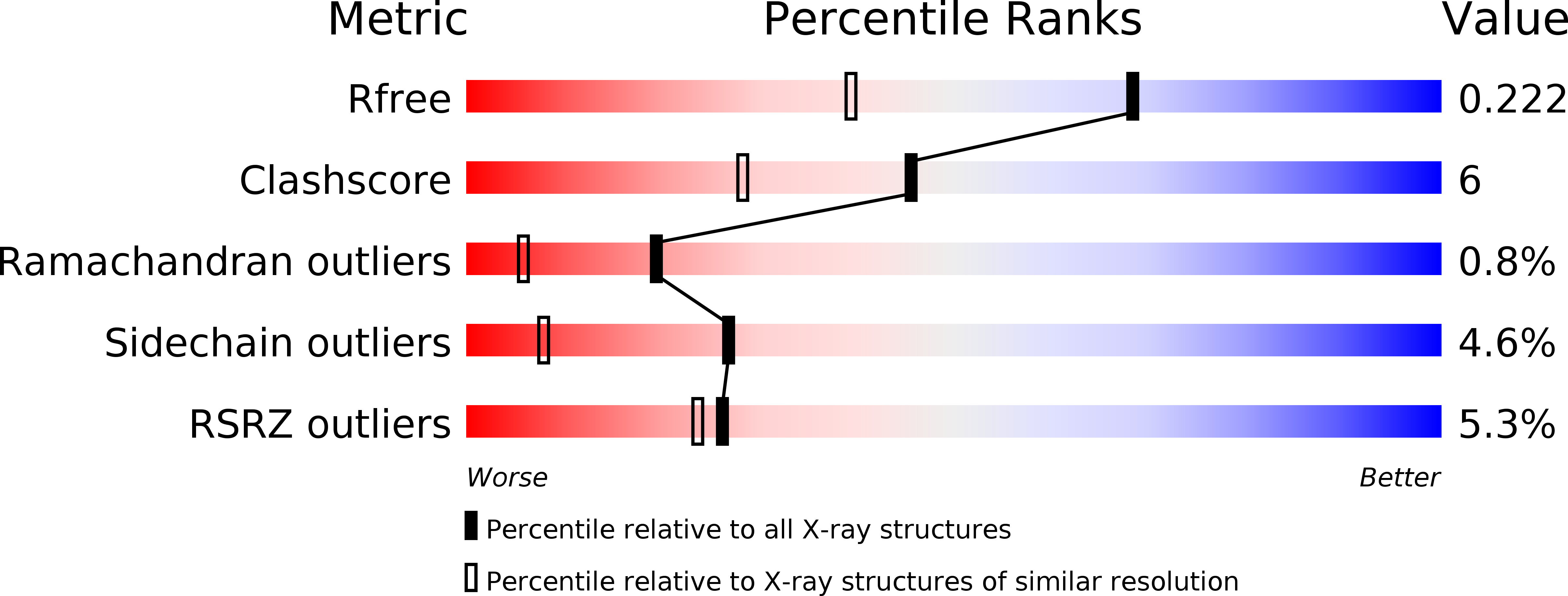
wwPDB Validation   3D Report Full Report


Ligand Structure Quality Assessment 


This is version 2.0 of the entry. See complete history


Literature

Structural and Functional Studies of gamma-Carboxyglutamic Acid Domains of Factor VIIa and Activated Protein C: Role of Magnesium at Physiological Calcium.

Vadivel, K.Agah, S.Messer, A.S.Cascio, D.Bajaj, M.S.Krishnaswamy, S.Esmon, C.T.Padmanabhan, K.Bajaj, S.P.

(2013) J Mol Biol 425: 1961-1981

  • DOI: https://doi.org/10.1016/j.jmb.2013.02.017
  • Primary Citation of Related Structures:  
    3JTC

  • PubMed Abstract: 

    Crystal structures of factor (F) VIIa/soluble tissue factor (TF), obtained under high Mg(2+) (50mM Mg(2+)/5mM Ca(2+)), have three of seven Ca(2+) sites in the γ-carboxyglutamic acid (Gla) domain replaced by Mg(2+) at positions 1, 4, and 7. We now report structures under low Mg(2+) (2.5mM Mg(2+)/5mM Ca(2+)) as well as under high Ca(2+) (5mM Mg(2+)/45 mM Ca(2+)). Under low Mg(2+), four Ca(2+) and three Mg(2+) occupy the same positions as in high-Mg(2+) structures. Conversely, under low Mg(2+), reexamination of the structure of Gla domain of activated Protein C (APC) complexed with soluble endothelial Protein C receptor (sEPCR) has position 4 occupied by Ca(2+) and positions 1 and 7 by Mg(2+). Nonetheless, in direct binding experiments, Mg(2+) replaced three Ca(2+) sites in the unliganded Protein C or APC. Further, the high-Ca(2+) condition was necessary to replace Mg4 in the FVIIa/soluble TF structure. In biological studies, Mg(2+) enhanced phospholipid binding to FVIIa and APC at physiological Ca(2+). Additionally, Mg(2+) potentiated phospholipid-dependent activations of FIX and FX by FVIIa/TF and inactivation of activated factor V by APC. Since APC and FVIIa bind to sEPCR involving similar interactions, we conclude that under the low-Mg(2+) condition, sEPCR binding to APC-Gla (or FVIIa-Gla) replaces Mg4 by Ca4 with an attendant conformational change in the Gla domain ω-loop. Moreover, since phospholipid and sEPCR bind to FVIIa or APC via the ω-loop, we predict that phospholipid binding also induces the functional Ca4 conformation in this loop. Cumulatively, the data illustrate that Mg(2+) and Ca(2+) act in concert to promote coagulation and anticoagulation.


  • Organizational Affiliation

    UCLA/Orthopaedic Hospital Department of Orthopaedic Surgery, University of California, Los Angeles, CA 90095, USA.


Macromolecules
Find similar proteins by:  (by identity cutoff)  |  3D Structure
Entity ID: 1
MoleculeChains Sequence LengthOrganismDetailsImage
Endothelial protein C receptor
A, B
193Homo sapiensMutation(s): 0 
UniProt & NIH Common Fund Data Resources
Find proteins for Q9UNN8 (Homo sapiens)
Explore Q9UNN8 
Go to UniProtKB:  Q9UNN8
PHAROS:  Q9UNN8
GTEx:  ENSG00000101000 
Entity Groups  
Sequence Clusters30% Identity50% Identity70% Identity90% Identity95% Identity100% Identity
UniProt GroupQ9UNN8
Glycosylation
Glycosylation Sites: 3Go to GlyGen: Q9UNN8-1
Sequence Annotations
Expand
  • Reference Sequence
Find similar proteins by:  (by identity cutoff)  |  3D Structure
Entity ID: 2
MoleculeChains Sequence LengthOrganismDetailsImage
Vitamin K-dependent protein C
C, D
33Homo sapiensMutation(s): 0 
EC: 3.4.21.69
UniProt & NIH Common Fund Data Resources
Find proteins for P04070 (Homo sapiens)
Explore P04070 
Go to UniProtKB:  P04070
PHAROS:  P04070
GTEx:  ENSG00000115718 
Entity Groups  
Sequence Clusters30% Identity50% Identity70% Identity90% Identity95% Identity100% Identity
UniProt GroupP04070
Sequence Annotations
Expand
  • Reference Sequence
Small Molecules
Ligands 4 Unique
IDChains Name / Formula / InChI Key2D Diagram3D Interactions
PTY
Query on PTY

Download Ideal Coordinates CCD File 
H [auth A],
L [auth B]
PHOSPHATIDYLETHANOLAMINE
C40 H80 N O8 P
NJGIRBISCGPRPF-KXQOOQHDSA-N
NAG
Query on NAG

Download Ideal Coordinates CCD File 
E [auth A]
F [auth A]
G [auth A]
I [auth B]
J [auth B]
E [auth A],
F [auth A],
G [auth A],
I [auth B],
J [auth B],
K [auth B]
2-acetamido-2-deoxy-beta-D-glucopyranose
C8 H15 N O6
OVRNDRQMDRJTHS-FMDGEEDCSA-N
CA
Query on CA

Download Ideal Coordinates CCD File 
N [auth C]
O [auth C]
P [auth C]
Q [auth C]
R [auth C]
N [auth C],
O [auth C],
P [auth C],
Q [auth C],
R [auth C],
U [auth D],
V [auth D],
W [auth D],
X [auth D],
Y [auth D]
CALCIUM ION
Ca
BHPQYMZQTOCNFJ-UHFFFAOYSA-N
MG
Query on MG

Download Ideal Coordinates CCD File 
M [auth C],
S [auth C],
T [auth D],
Z [auth D]
MAGNESIUM ION
Mg
JLVVSXFLKOJNIY-UHFFFAOYSA-N
Modified Residues  1 Unique
IDChains TypeFormula2D DiagramParent
CGU
Query on CGU
C, D
L-PEPTIDE LINKINGC6 H9 N O6GLU
Experimental Data & Validation

Experimental Data

  • Method: X-RAY DIFFRACTION
  • Resolution: 1.60 Å
  • R-Value Free: 0.226 
  • R-Value Work: 0.191 
  • R-Value Observed: 0.192 
  • Space Group: P 1 21 1
Unit Cell:
Length ( Å )Angle ( ˚ )
a = 59.22α = 90
b = 62.36β = 101.81
c = 71.03γ = 90
Software Package:
Software NamePurpose
REFMACrefinement
PDB_EXTRACTdata extraction
HKL-2000data reduction
SCALEPACKdata scaling

Structure Validation

View Full Validation Report


Ligand Structure Quality Assessment 


Entry History 

Deposition Data

Revision History  (Full details and data files)

  • Version 1.0: 2011-04-06
    Type: Initial release
  • Version 1.1: 2011-07-13
    Changes: Version format compliance
  • Version 1.2: 2013-03-20
    Changes: Database references
  • Version 1.3: 2013-07-03
    Changes: Database references
  • Version 2.0: 2020-07-29
    Type: Remediation
    Reason: Carbohydrate remediation
    Changes: Advisory, Atomic model, Data collection, Derived calculations, Non-polymer description, Structure summary